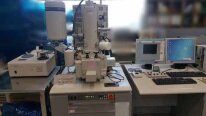
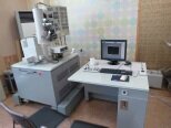
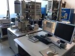
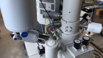
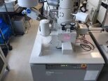
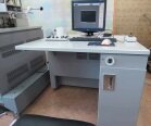
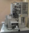
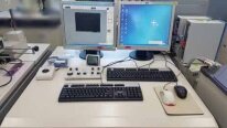
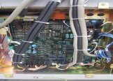
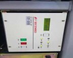
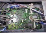
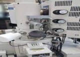
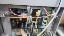
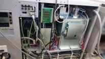
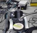
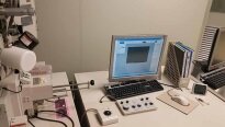
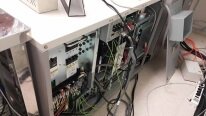
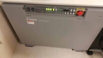
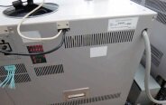
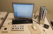
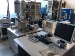
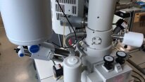
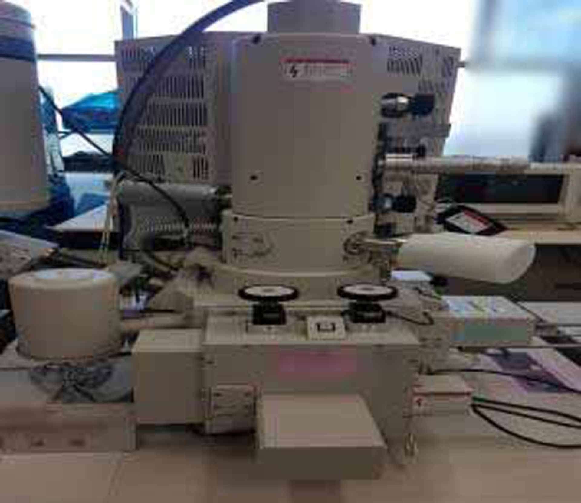
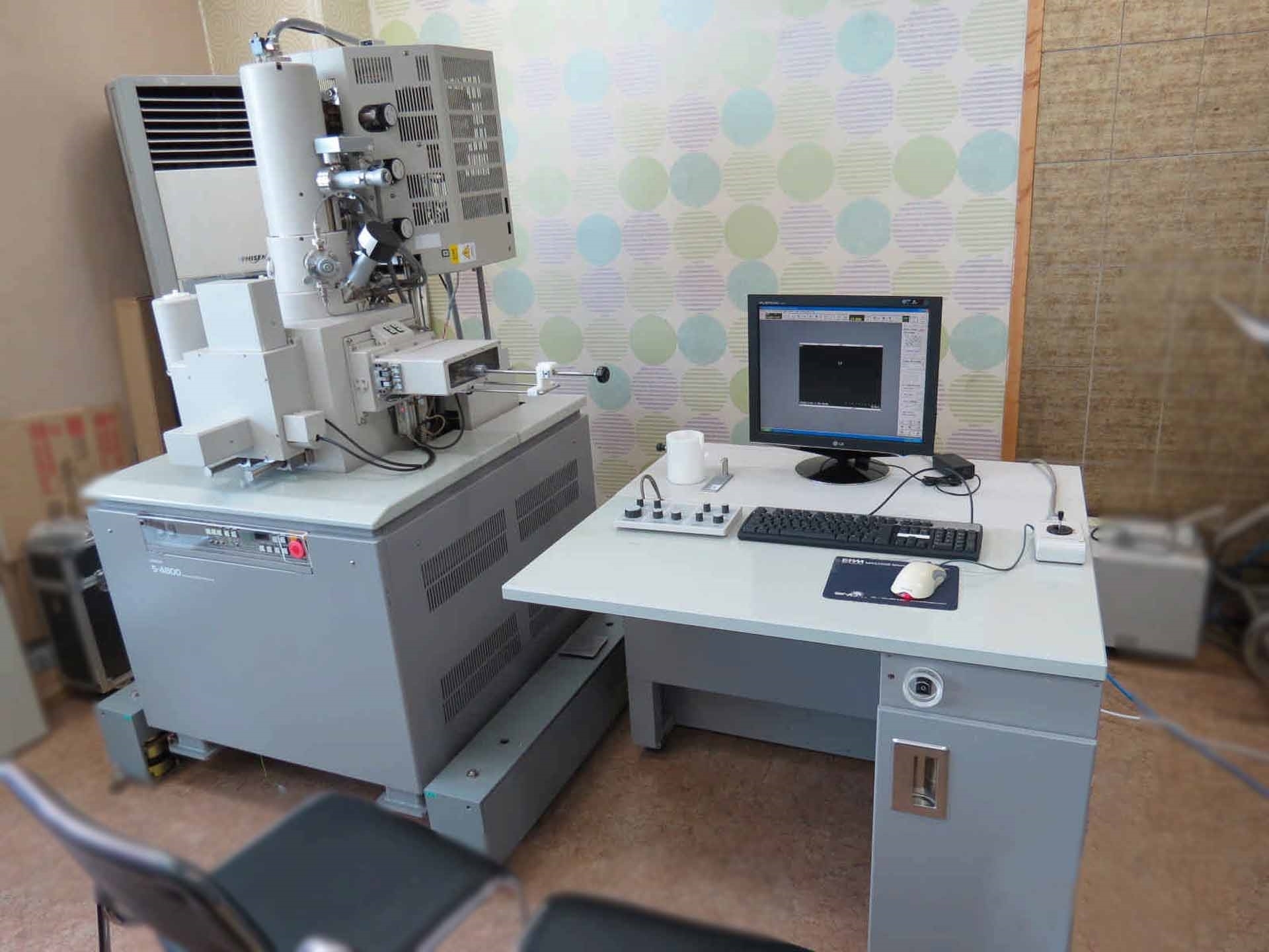
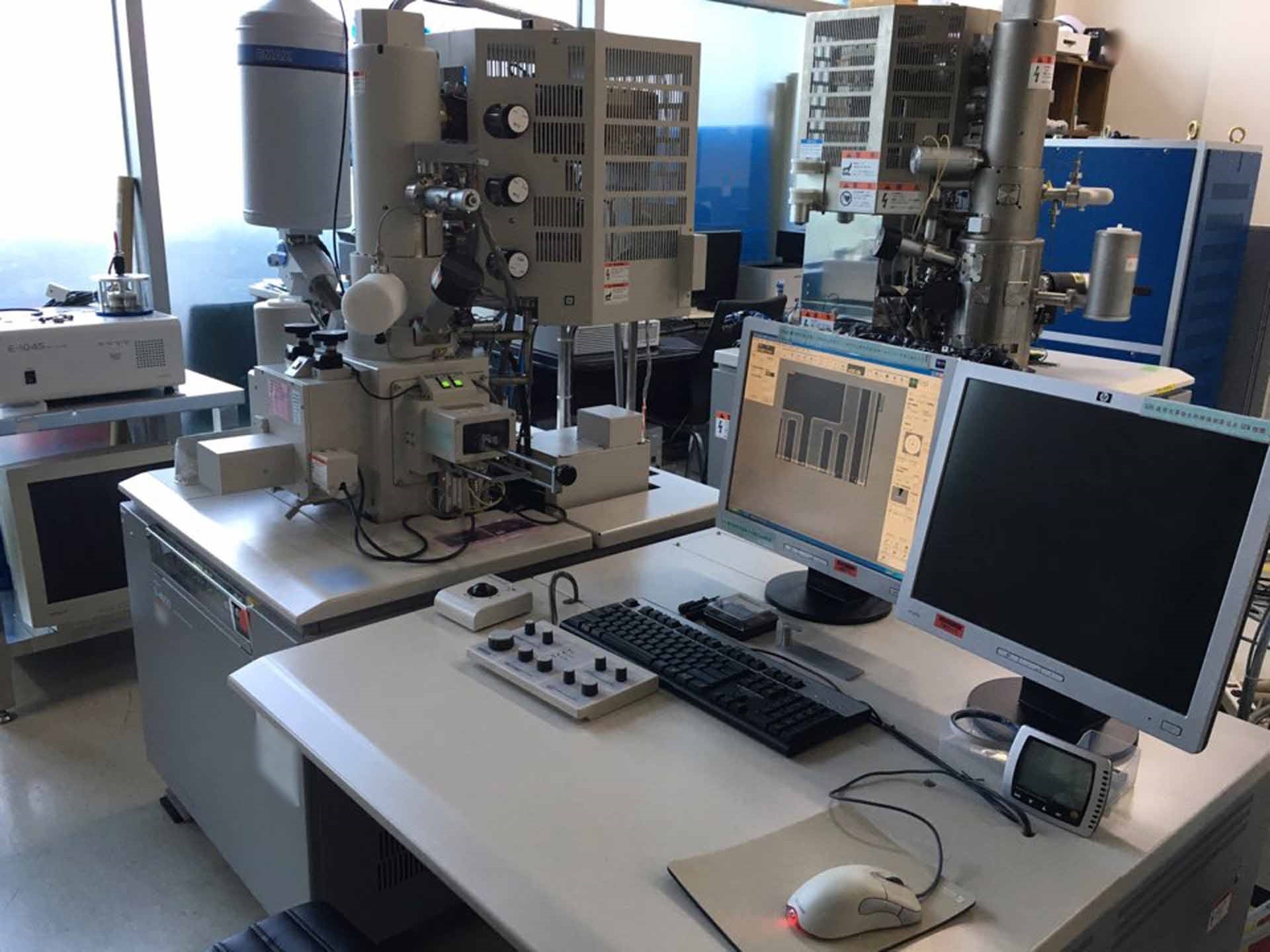
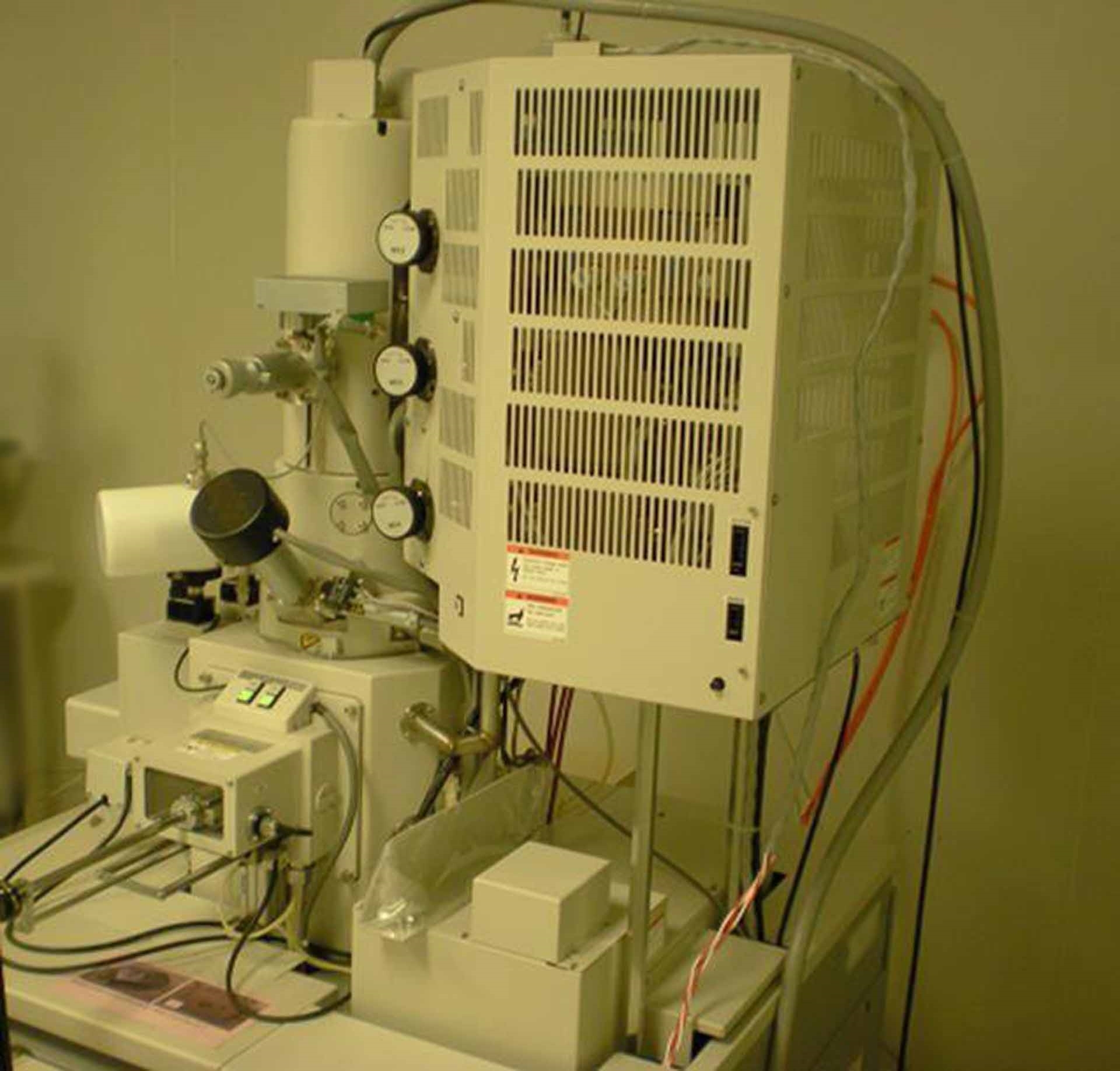
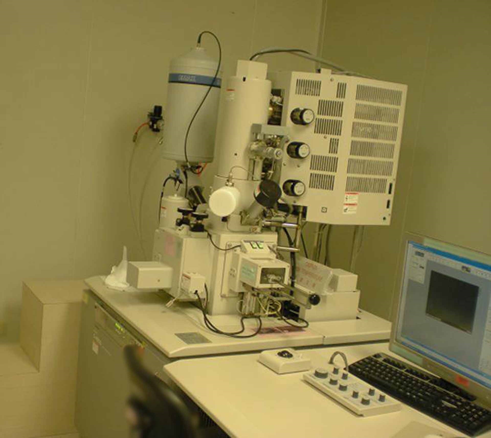
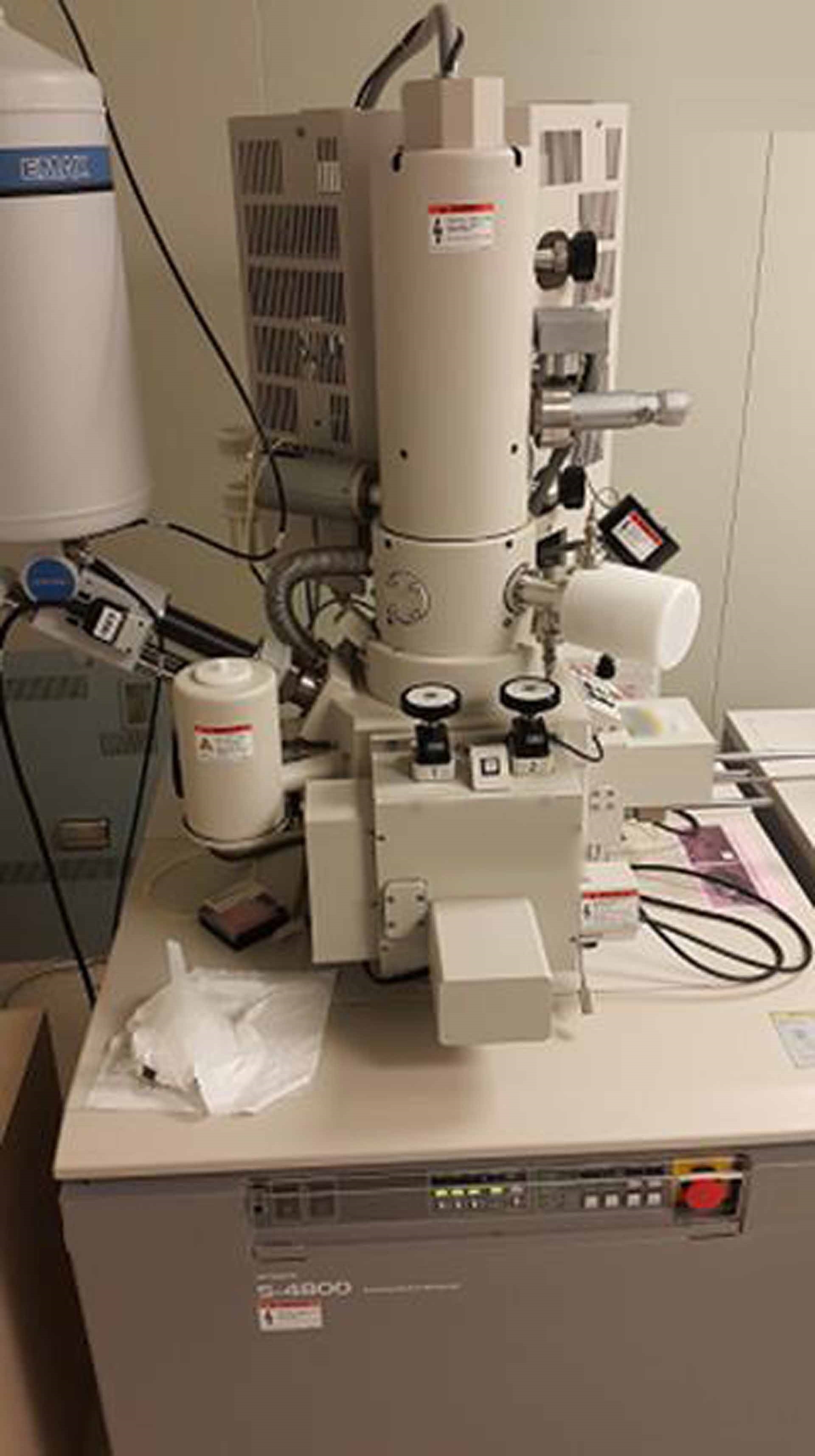
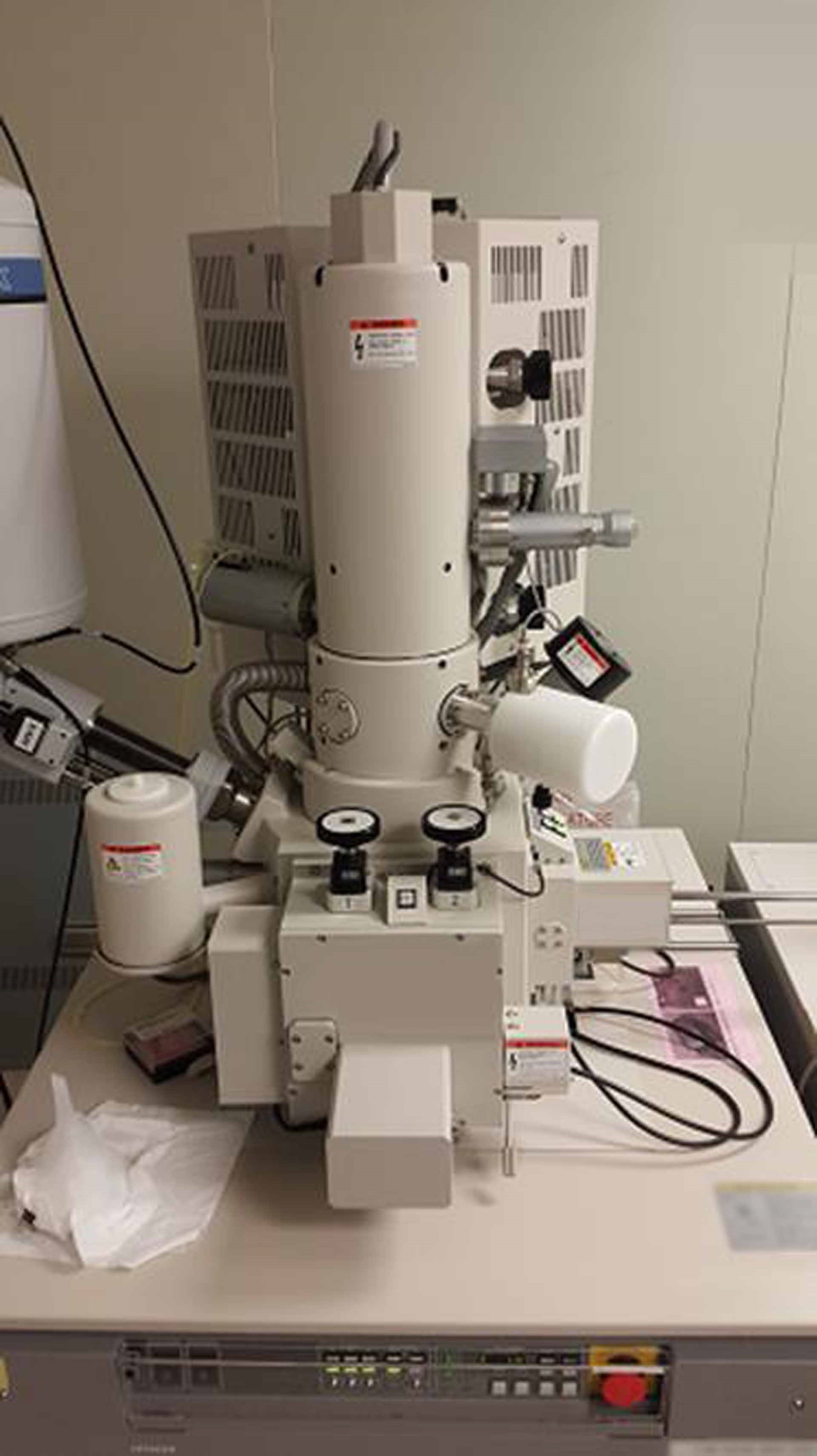
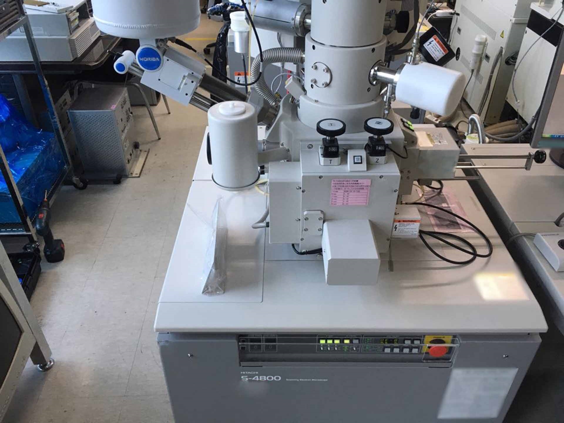
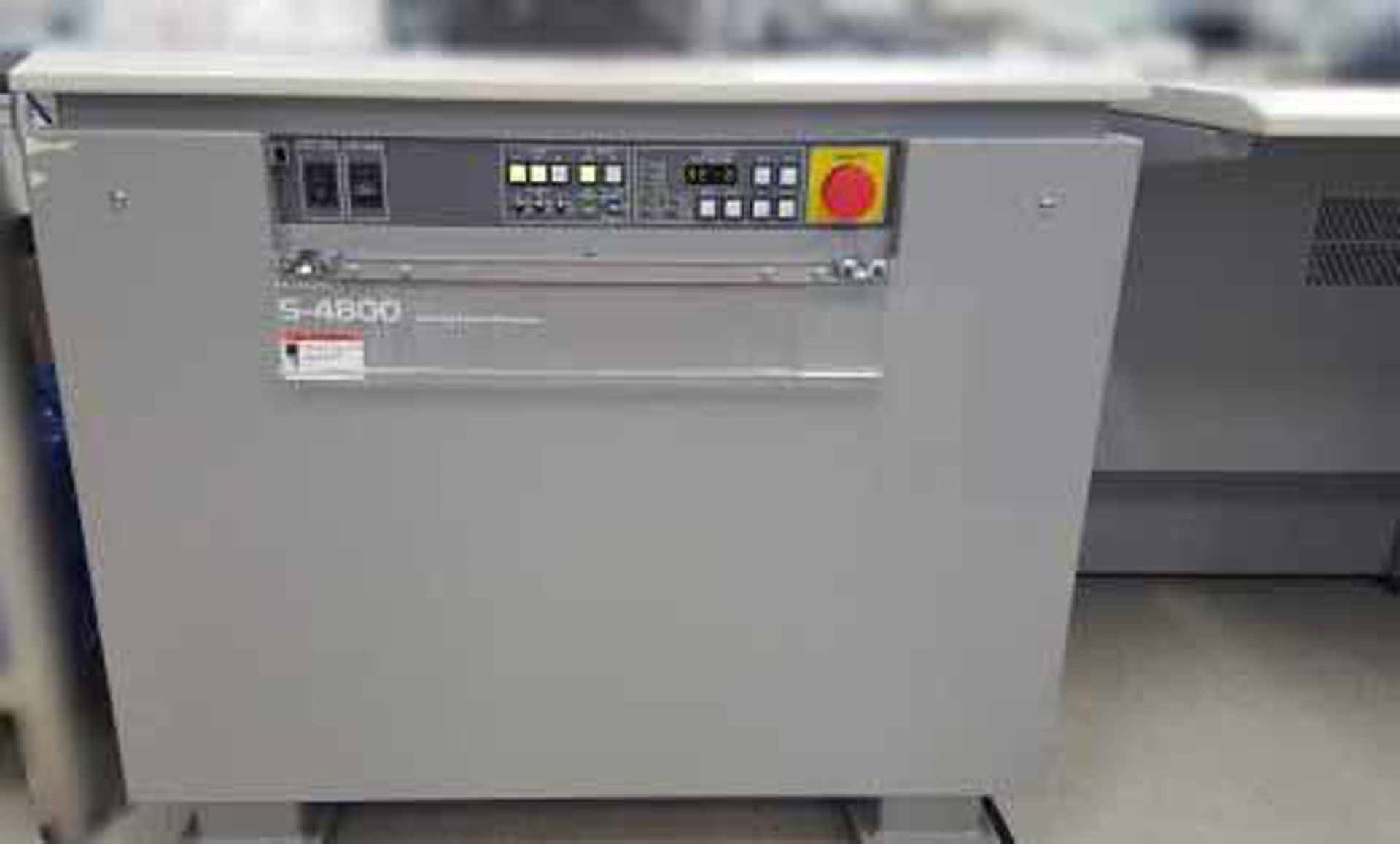
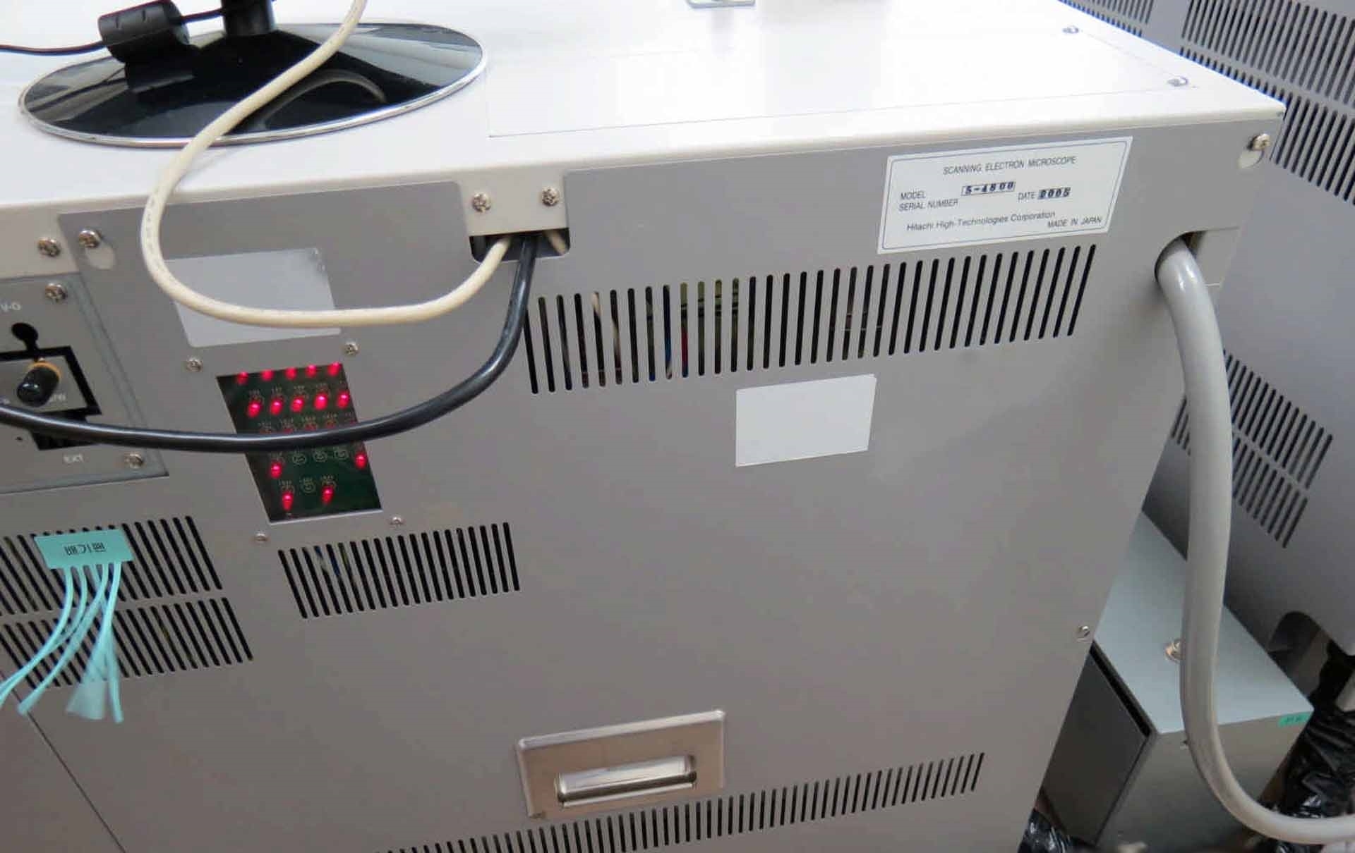
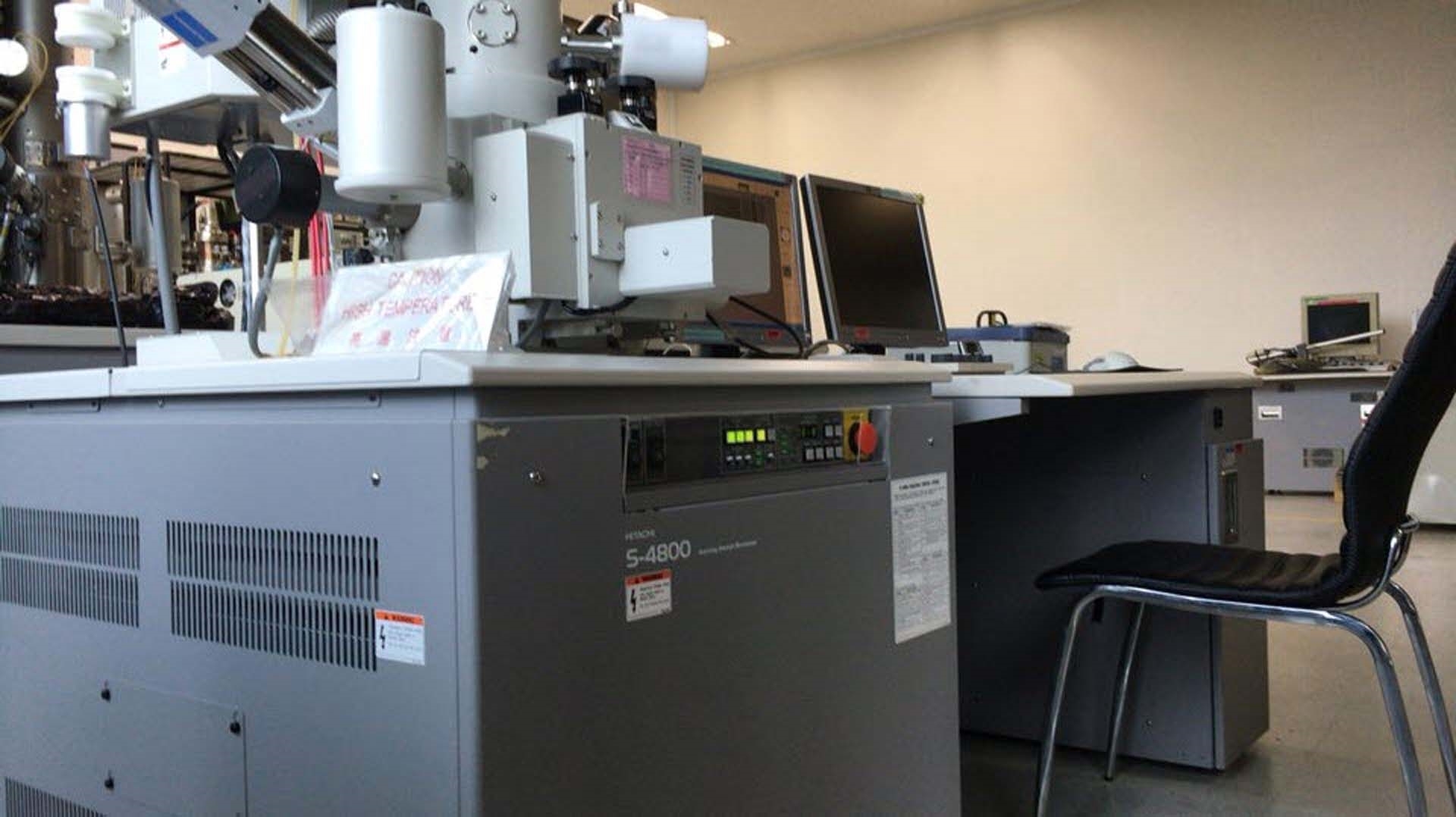
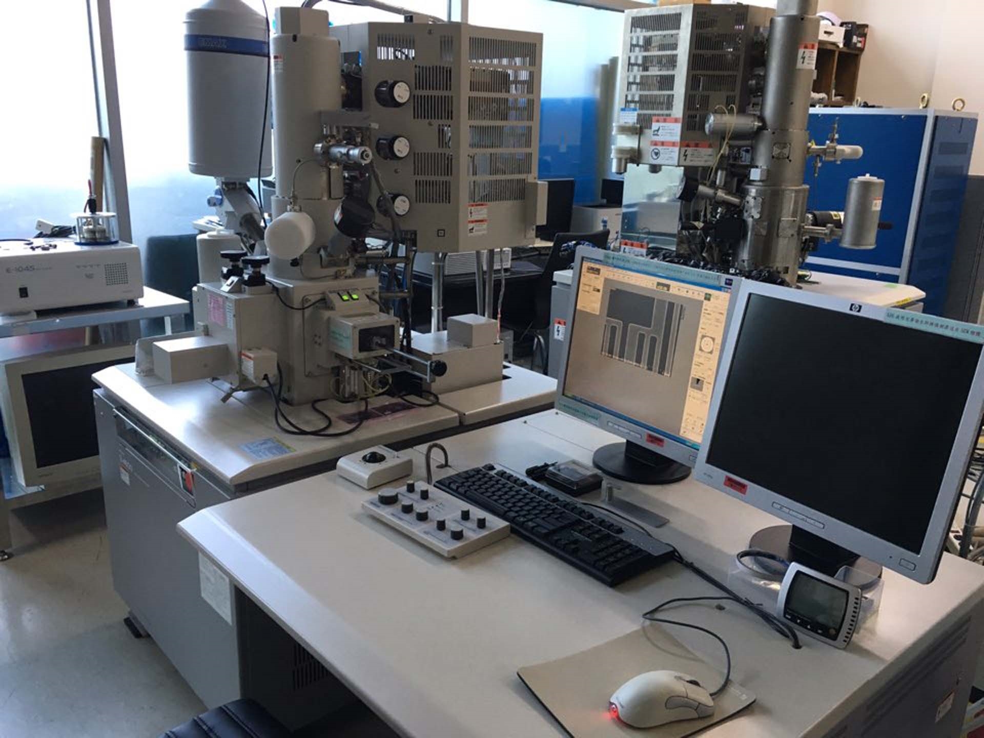
판매용 중고 HITACHI S-4800 #9209753
URL이 복사되었습니다!
확대하려면 누르십시오


















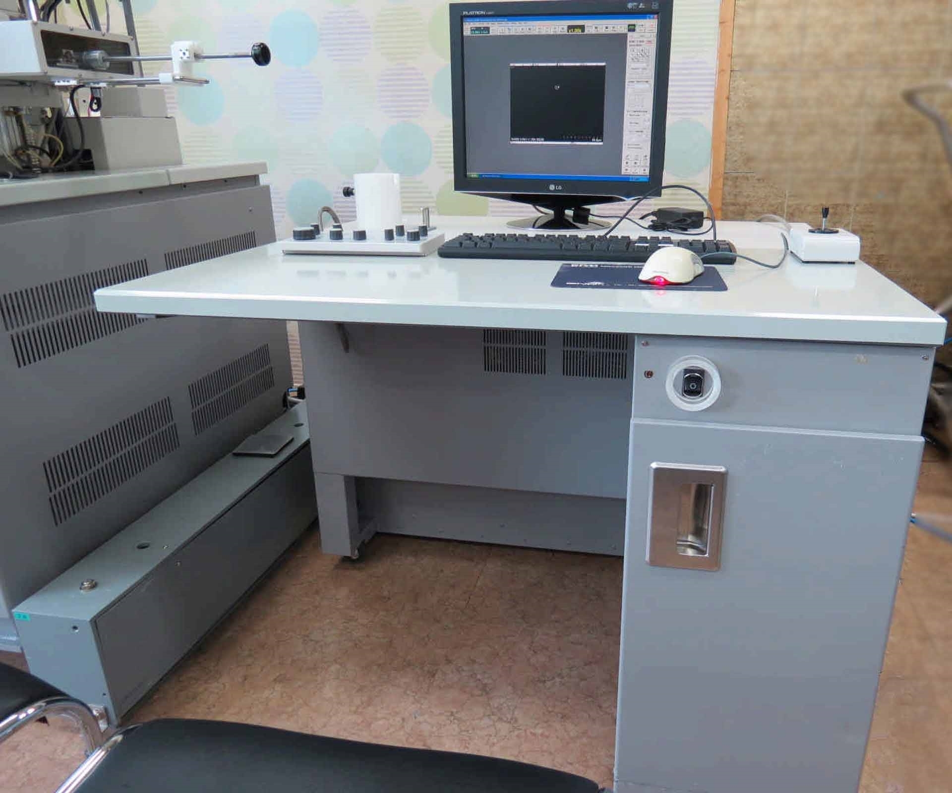

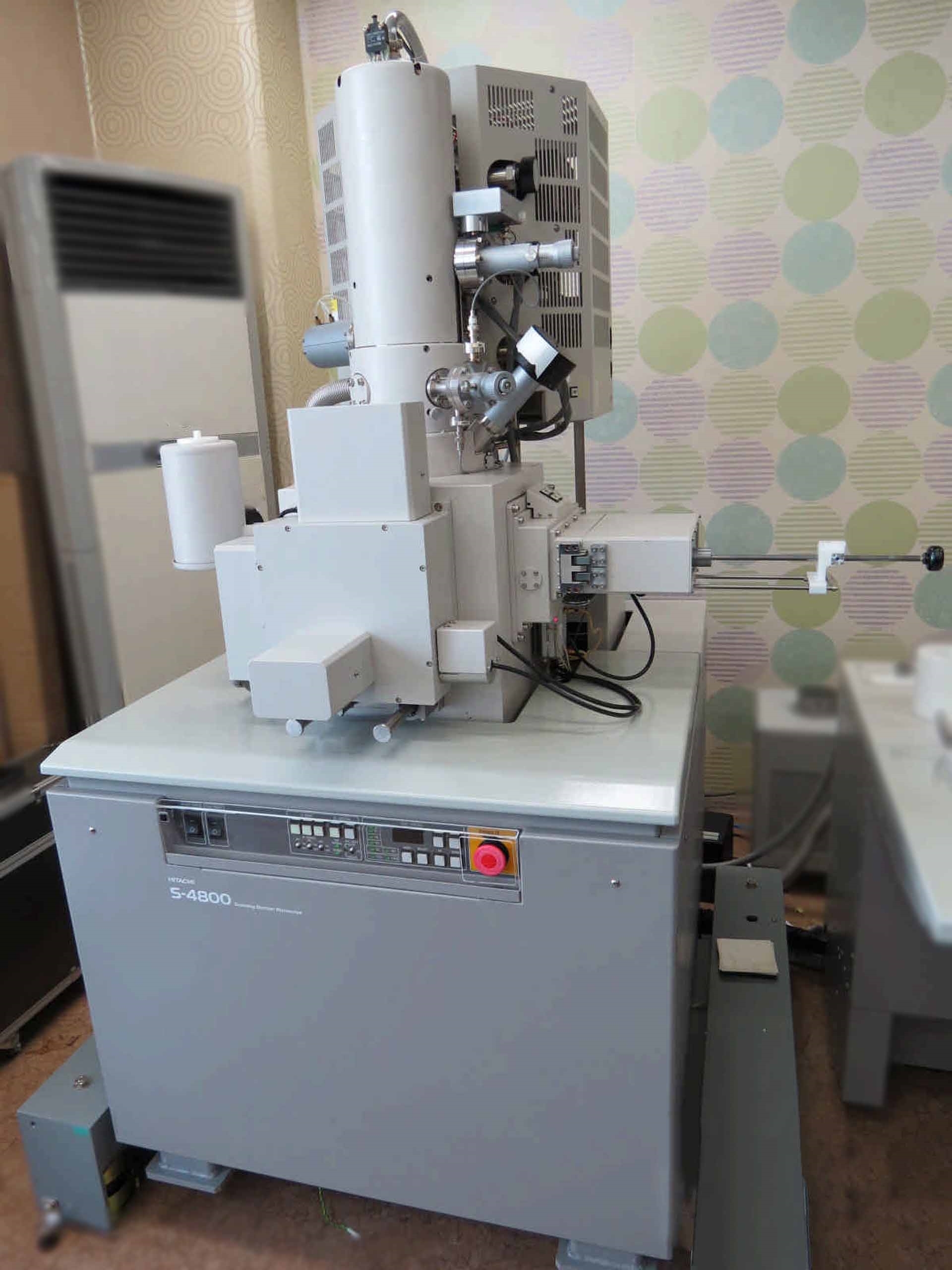

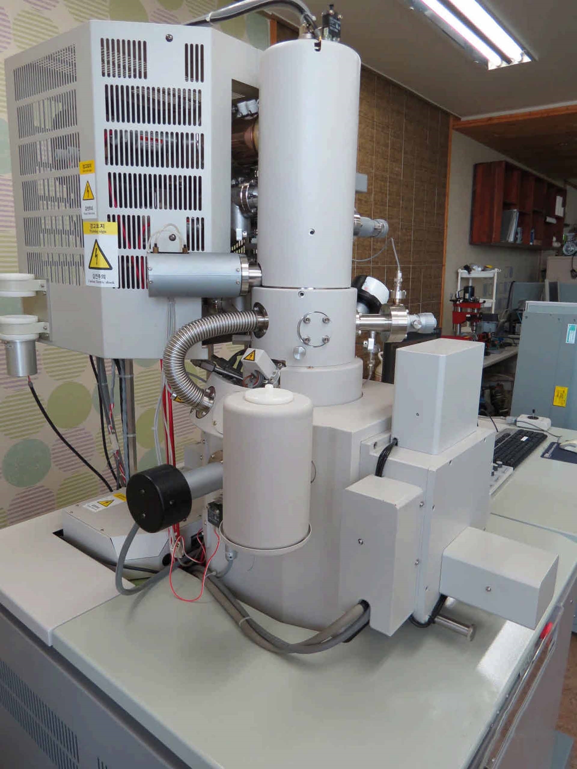

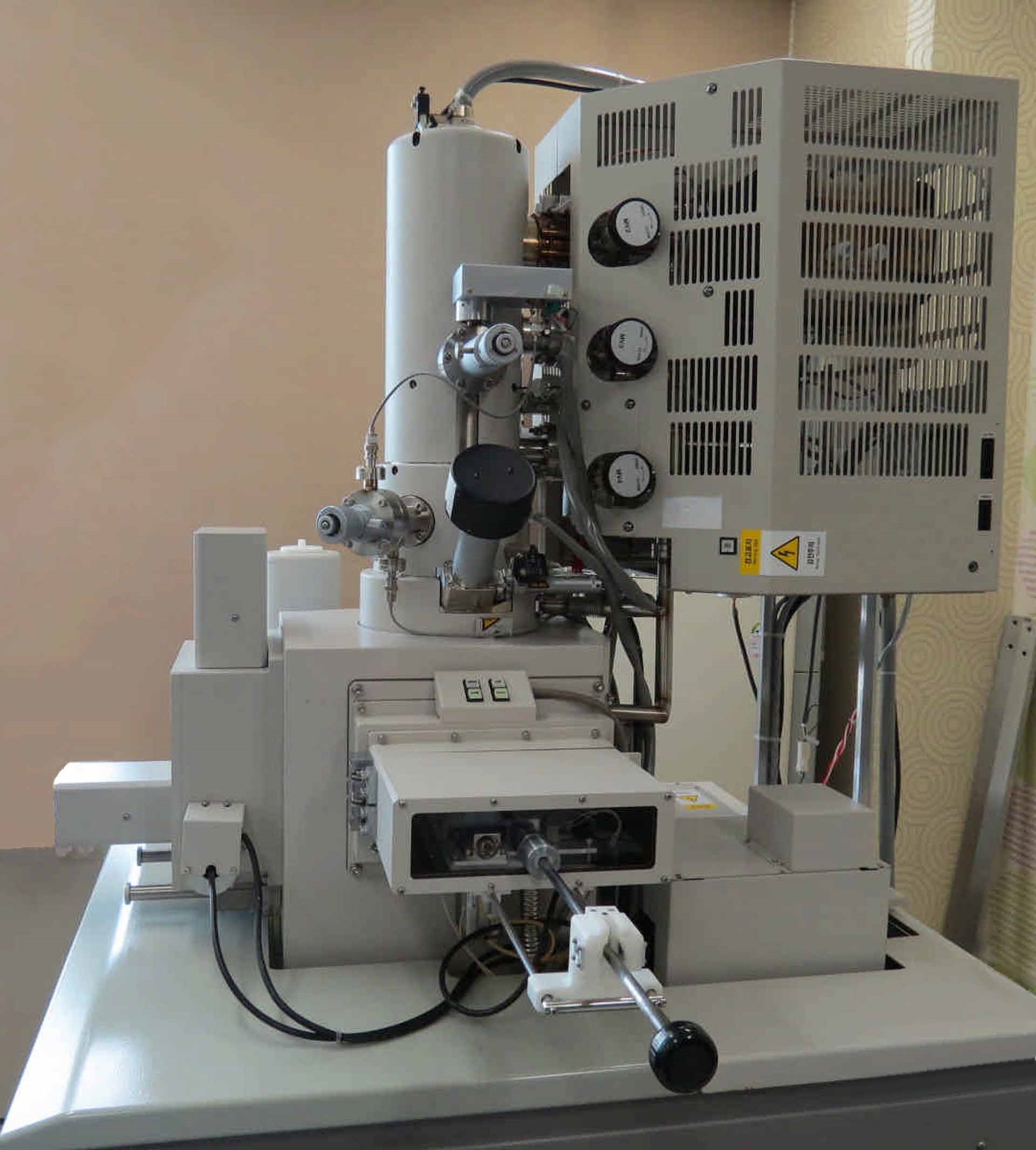





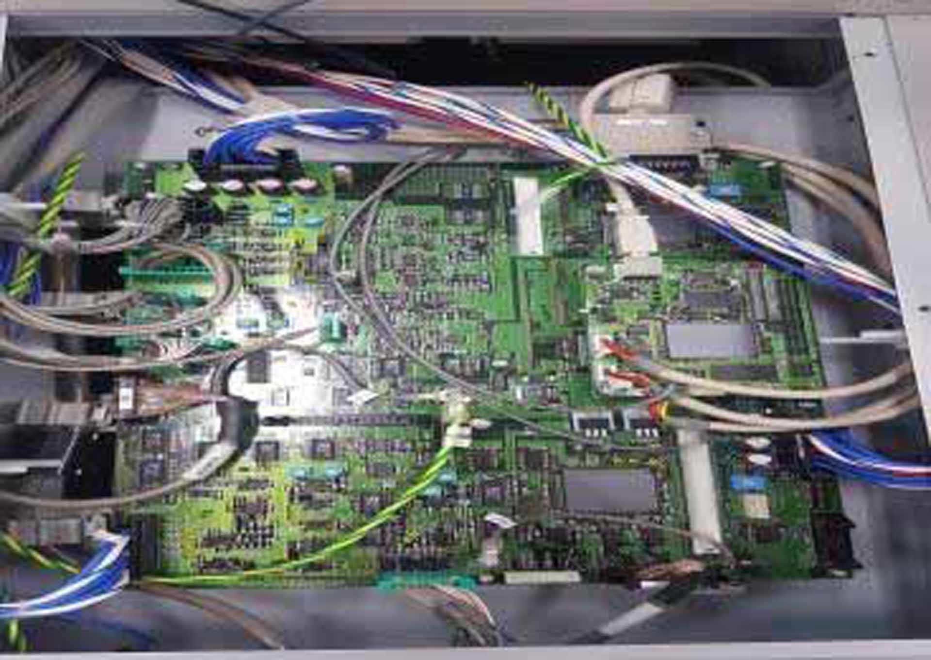

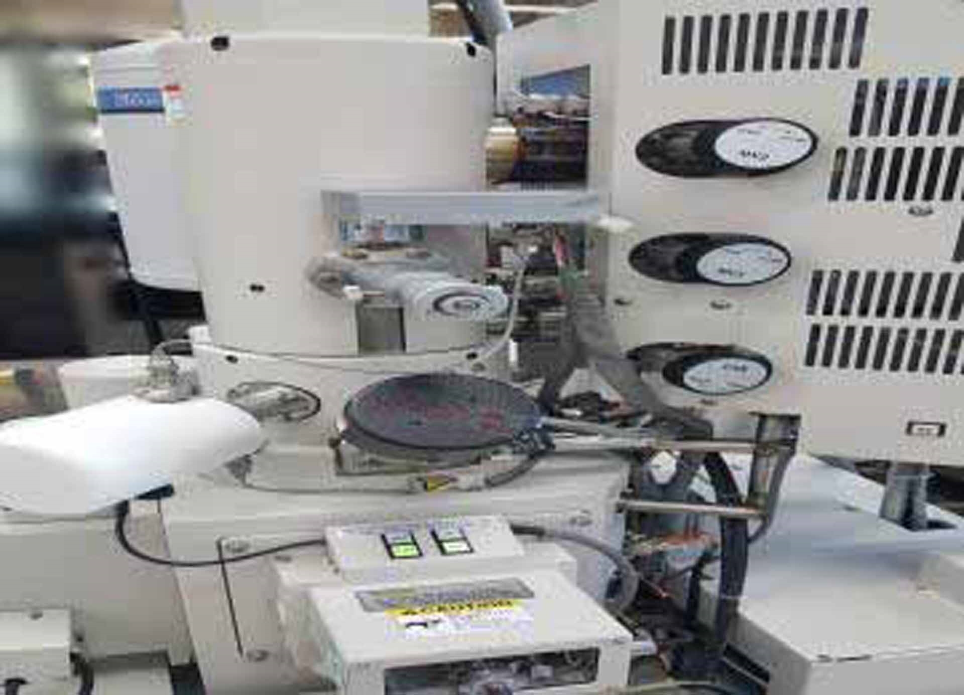

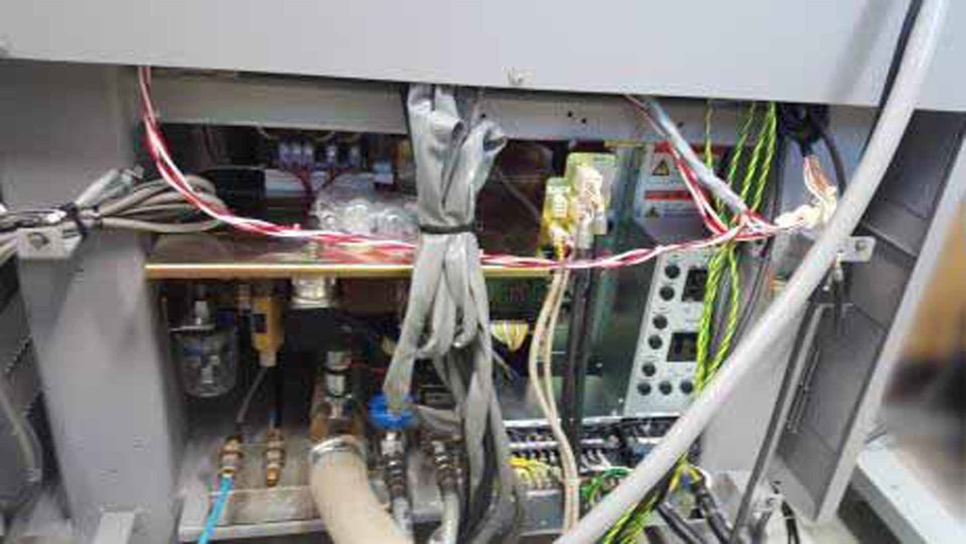

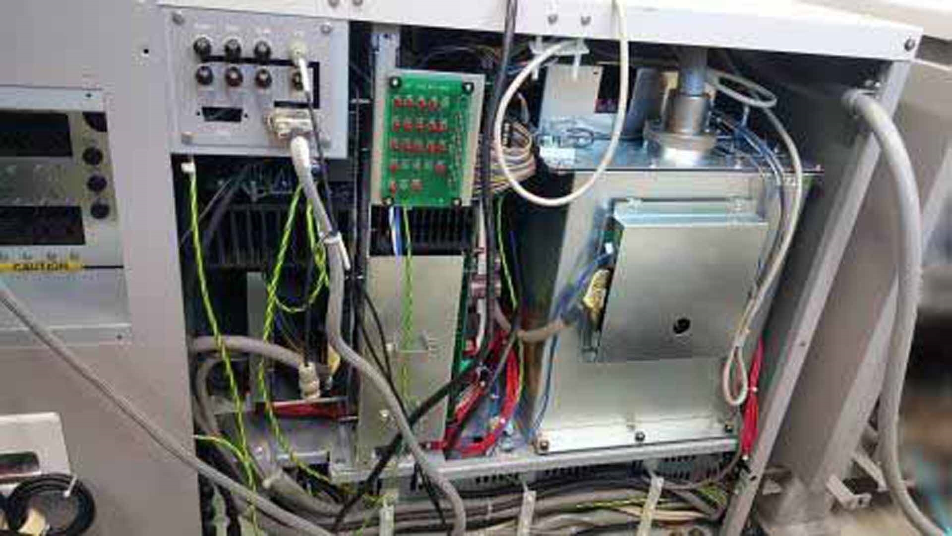

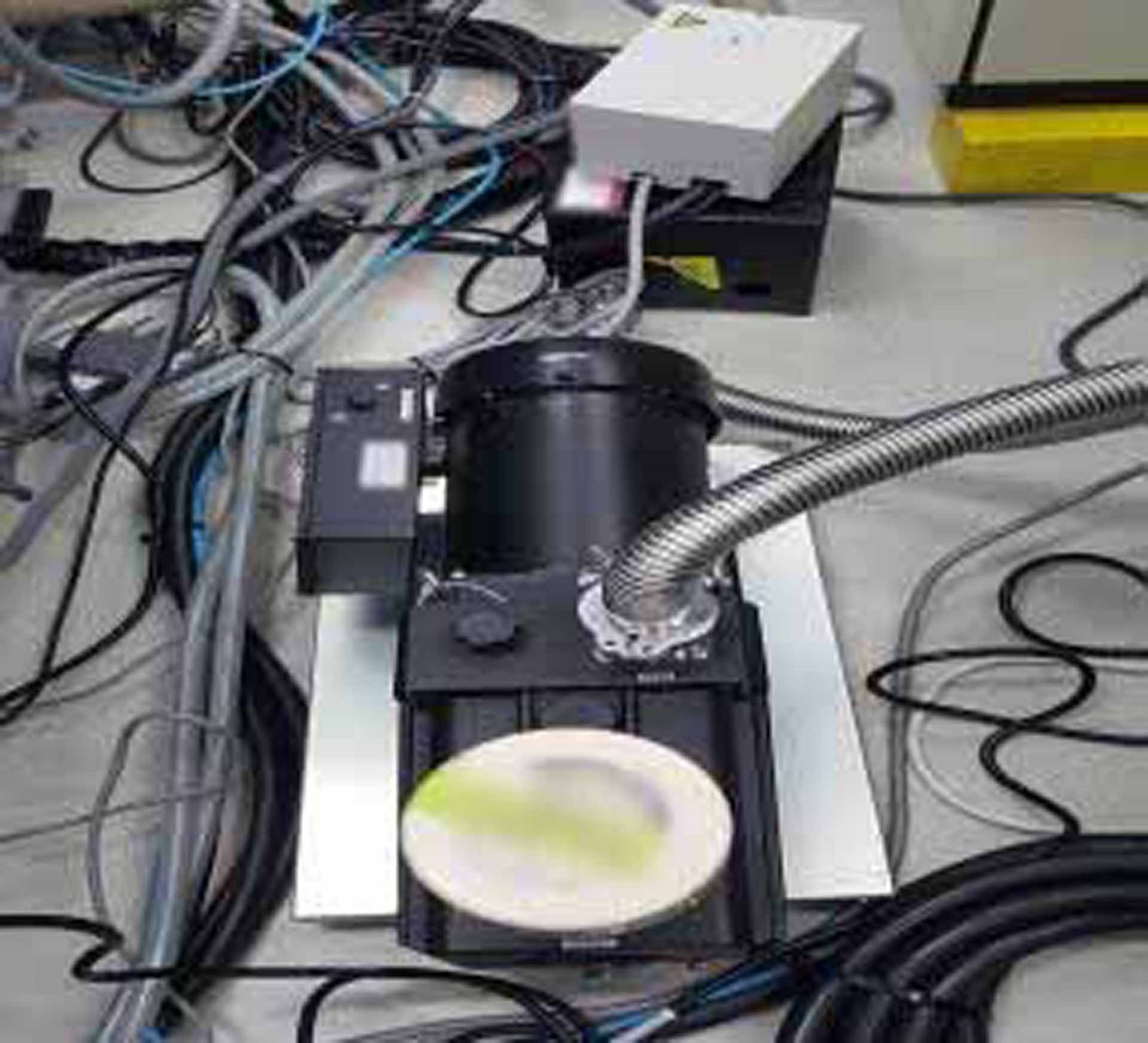




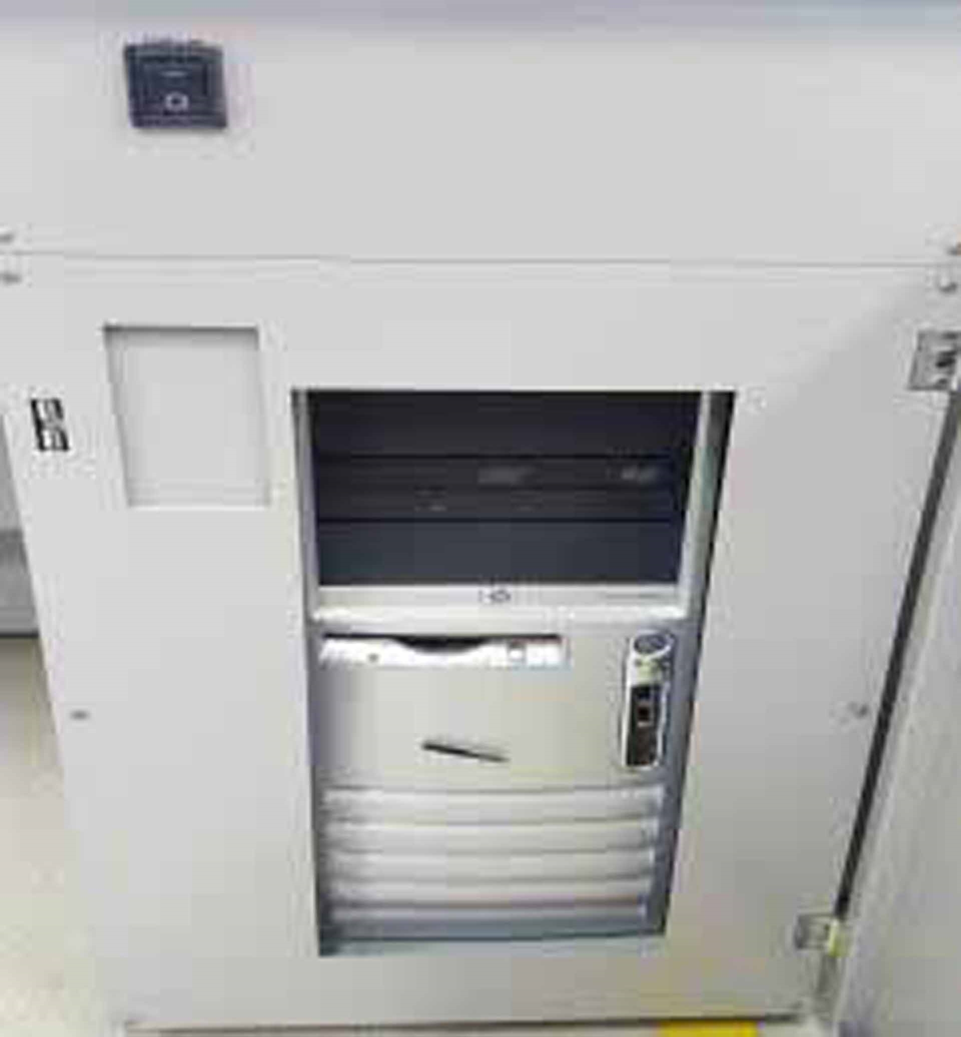

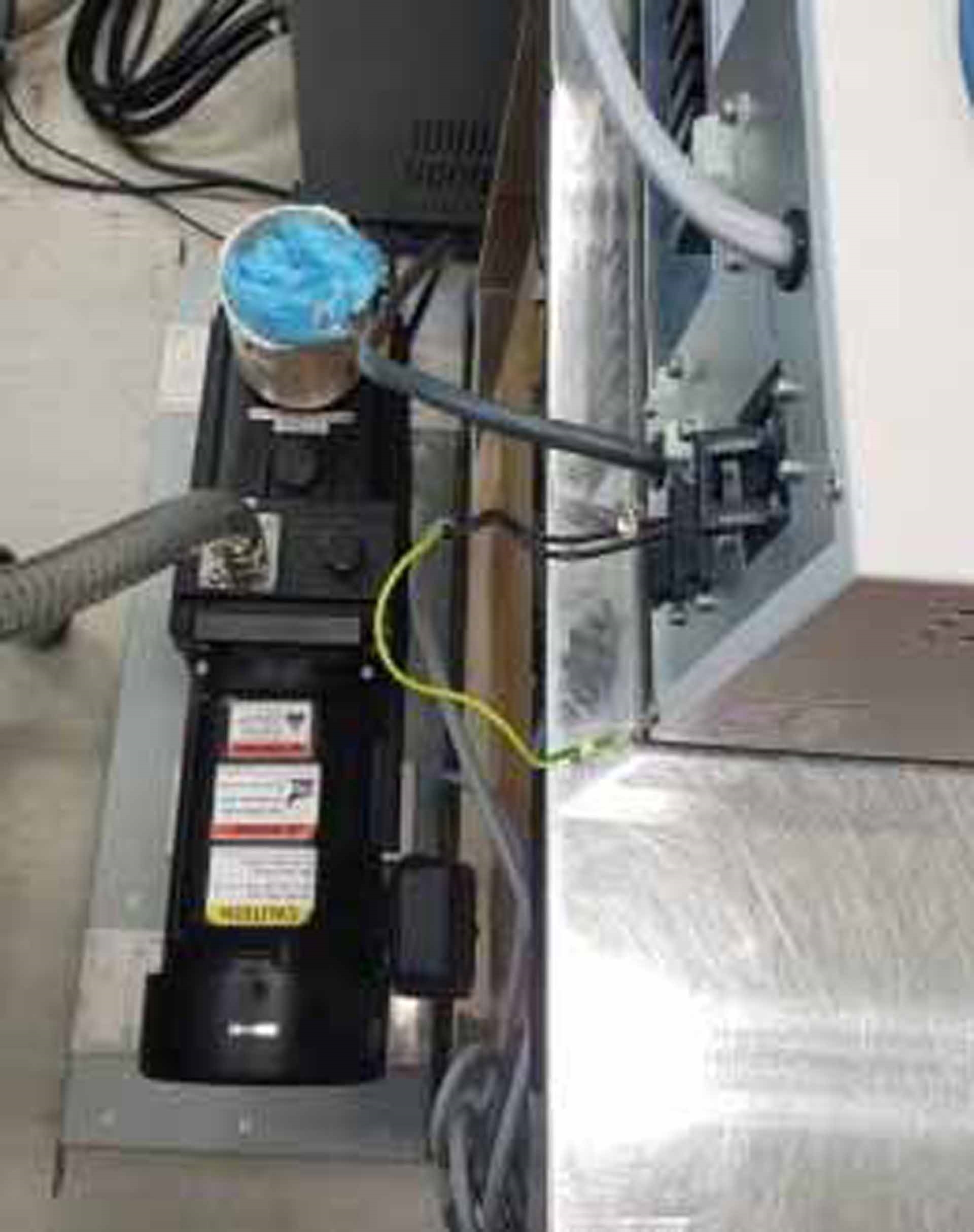

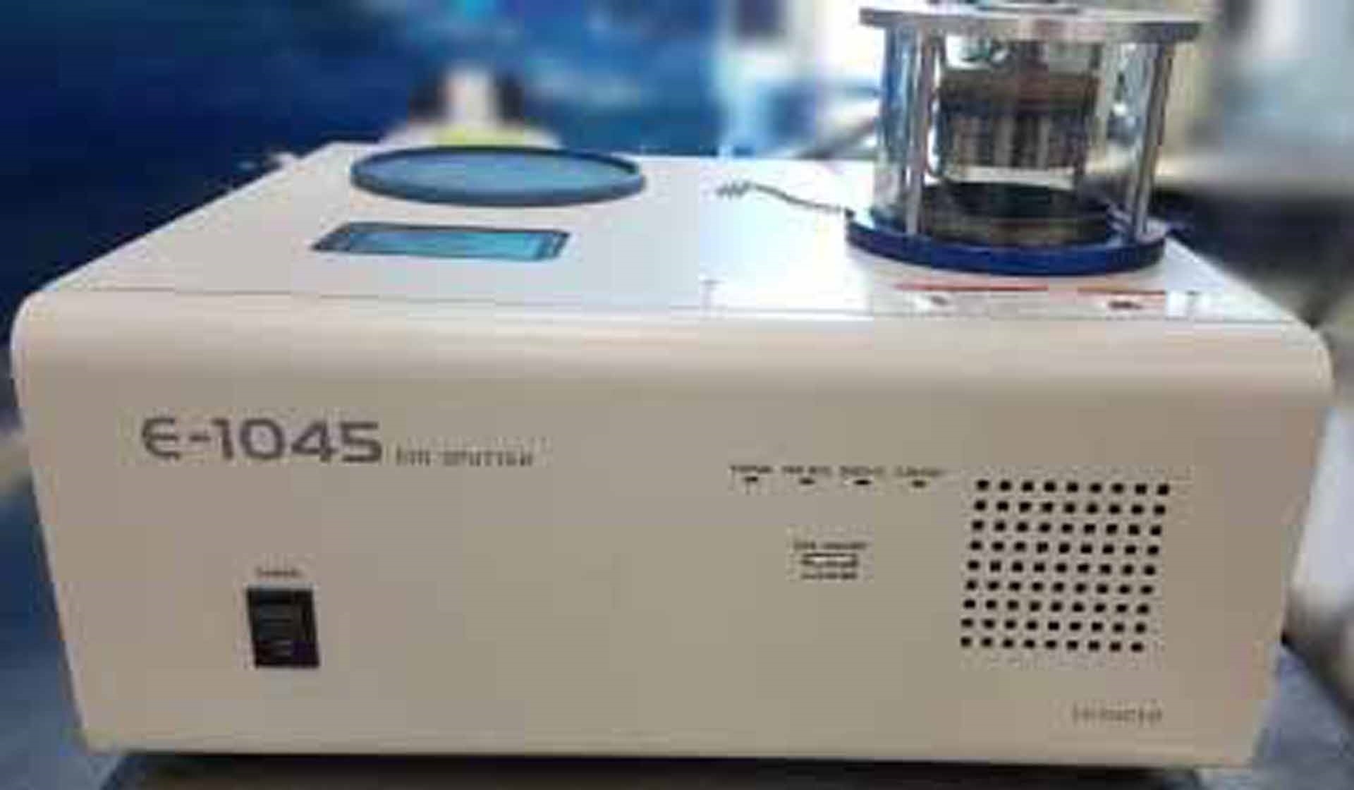

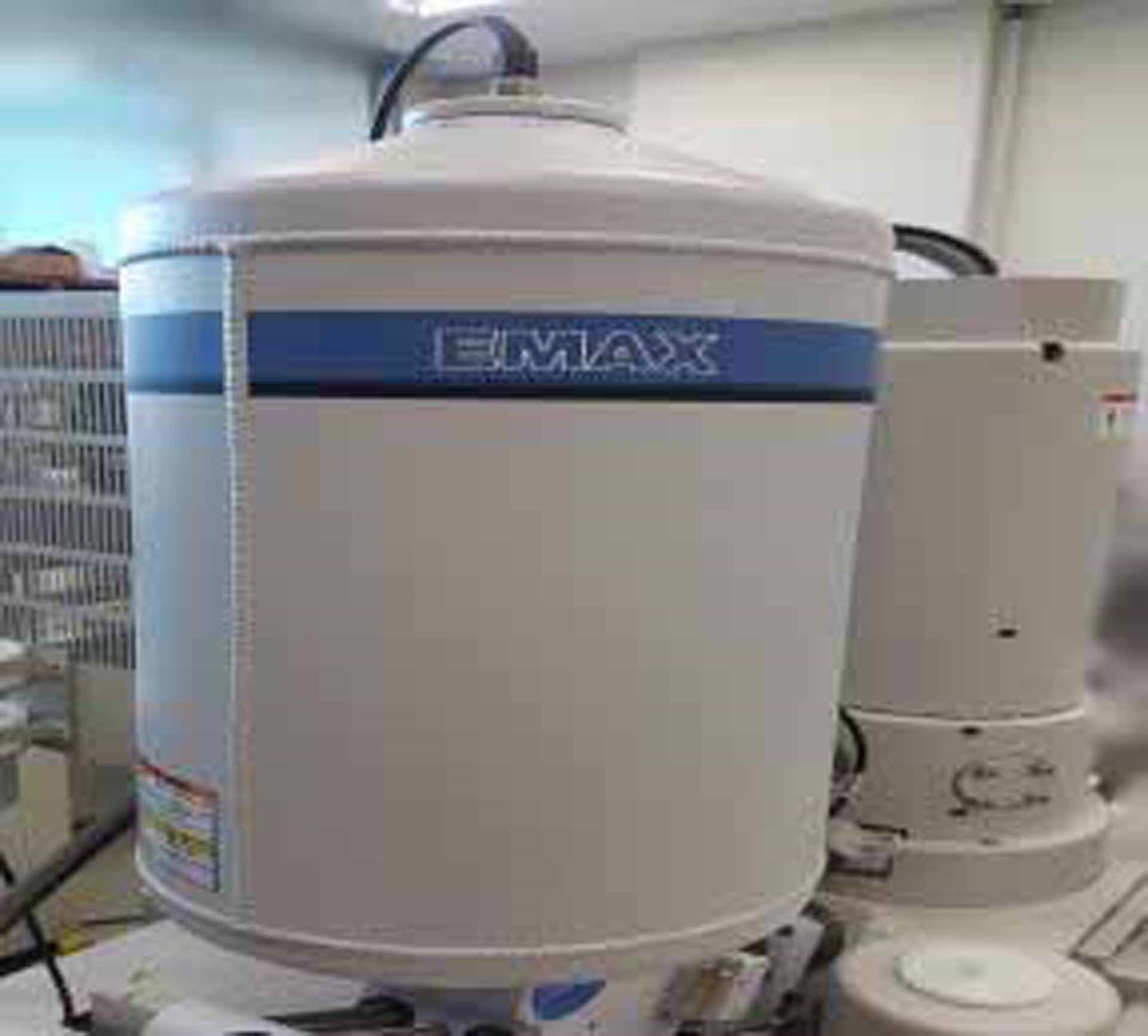





























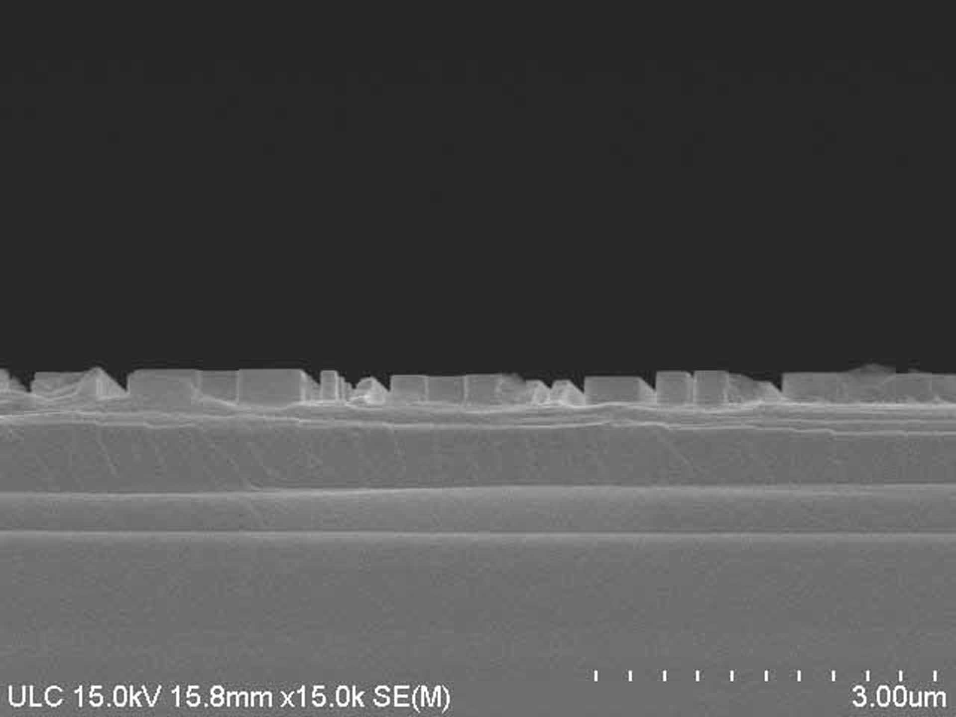

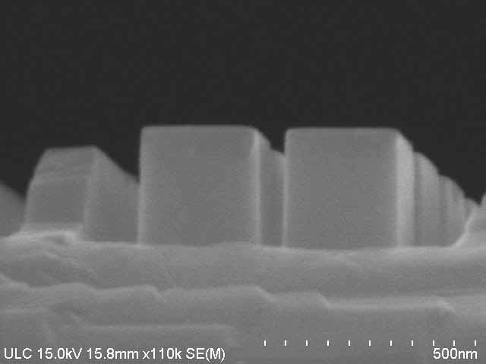

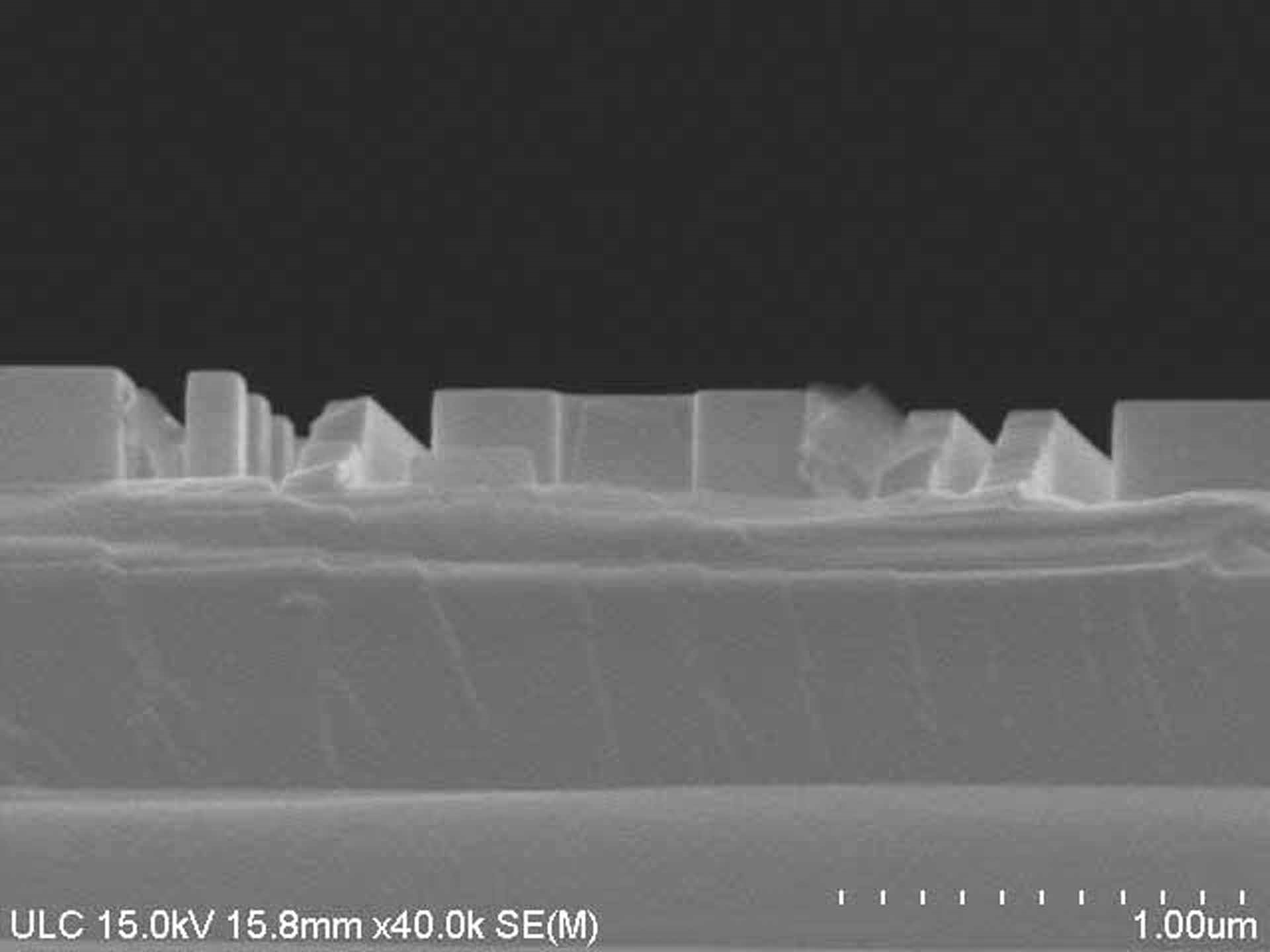

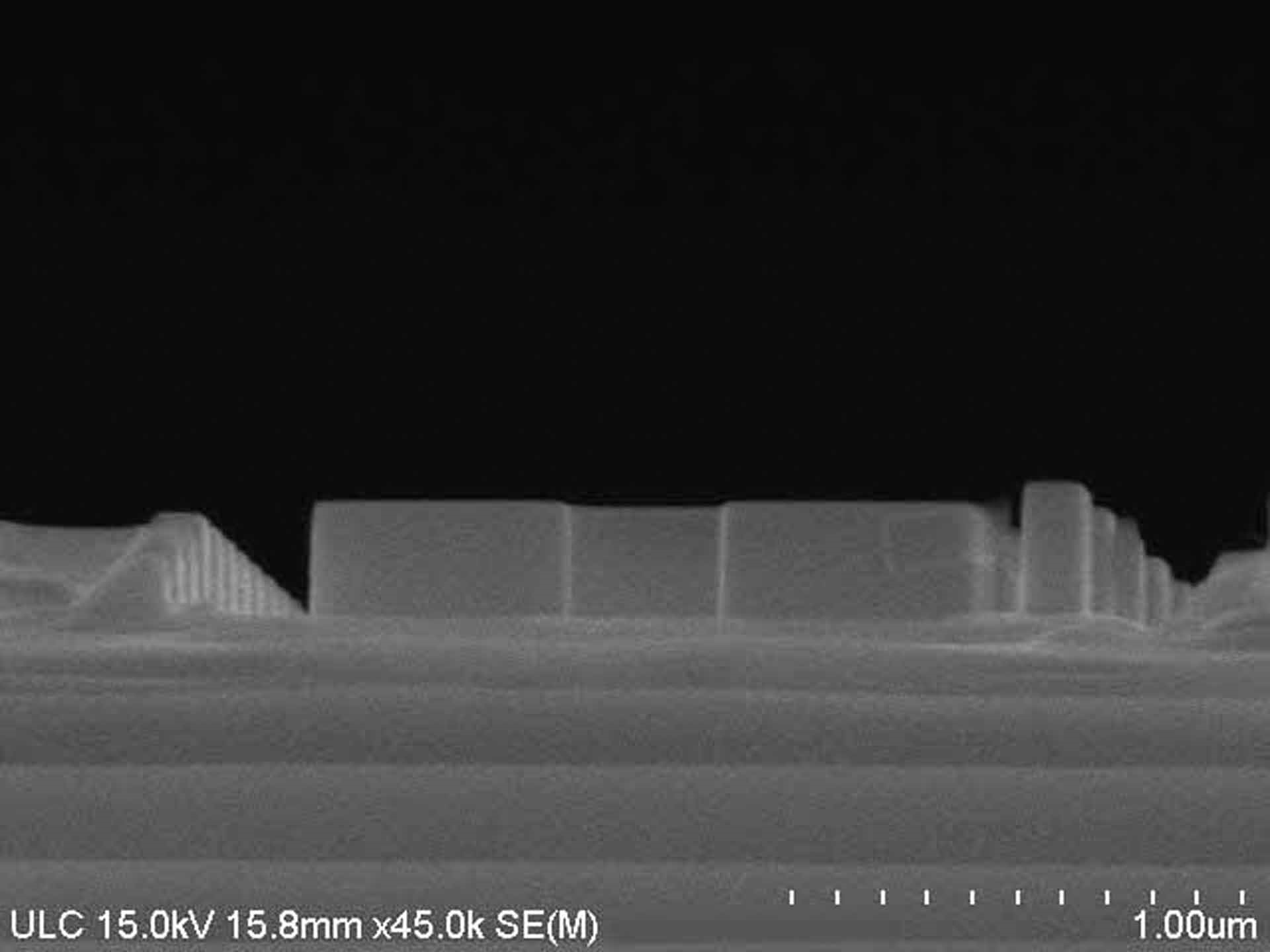































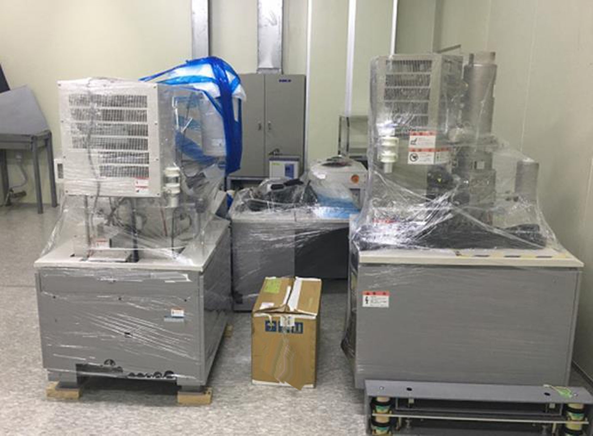

ID: 9209753
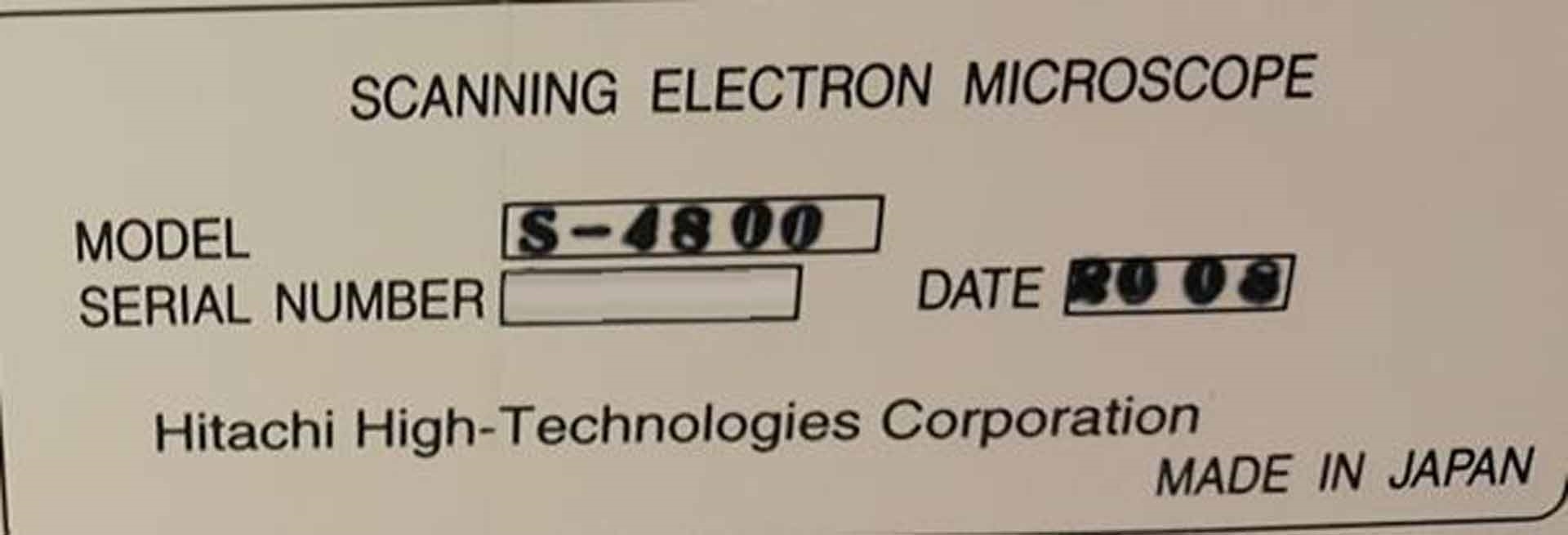
빈티지: 2008
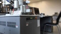
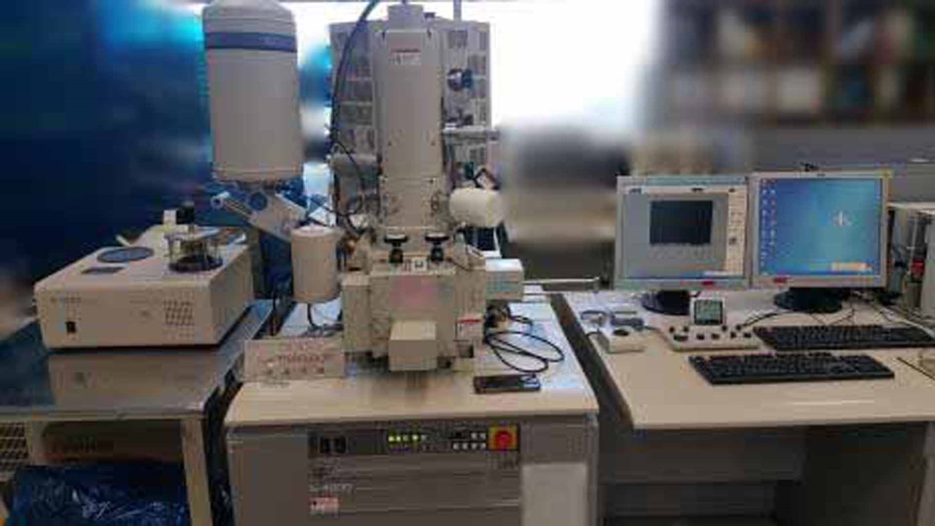
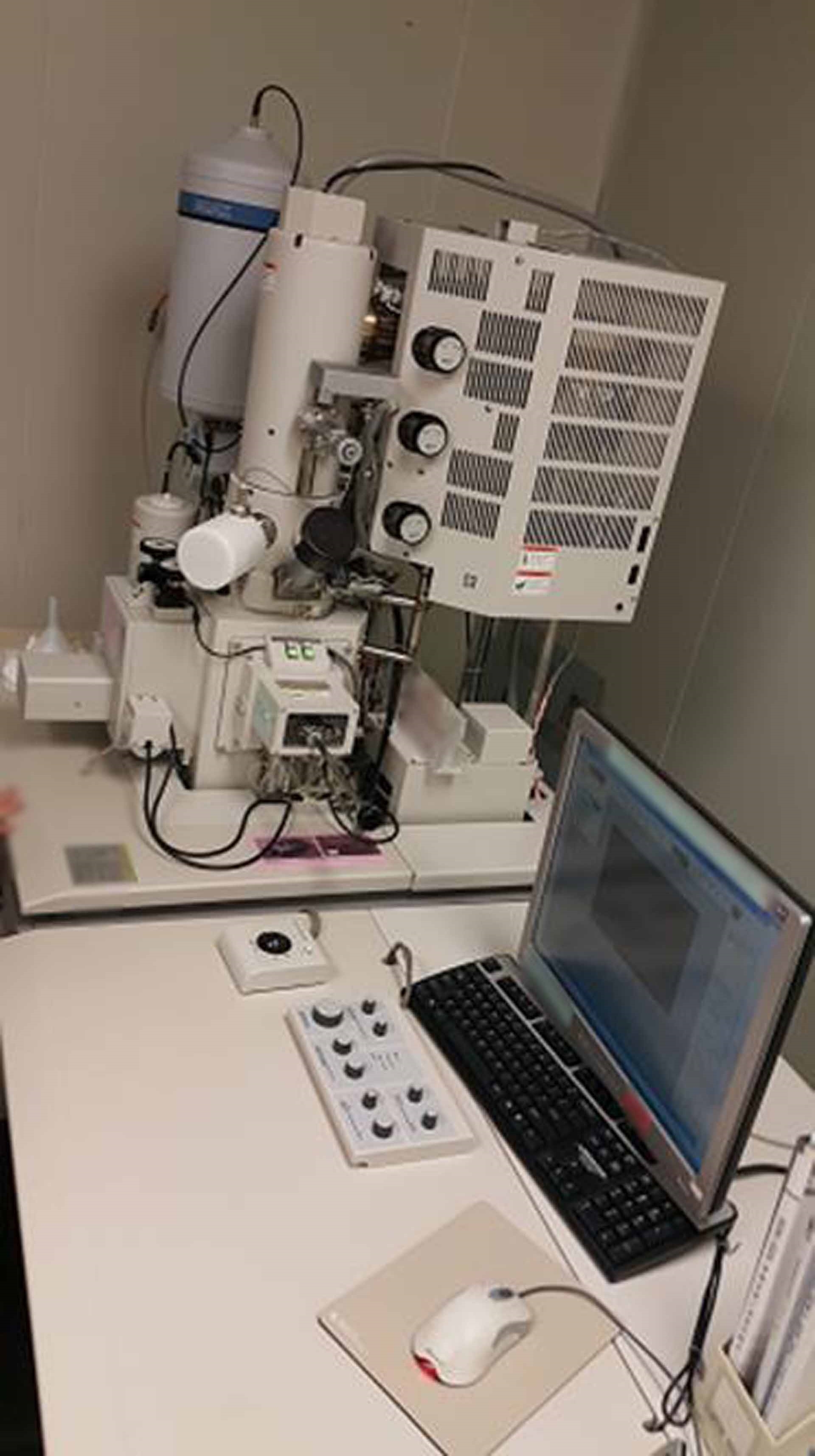
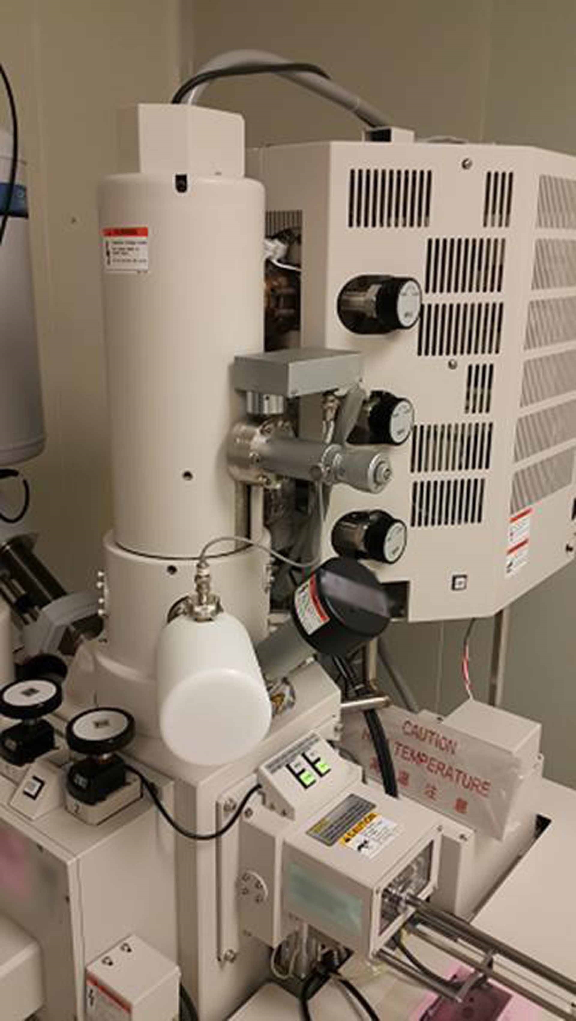
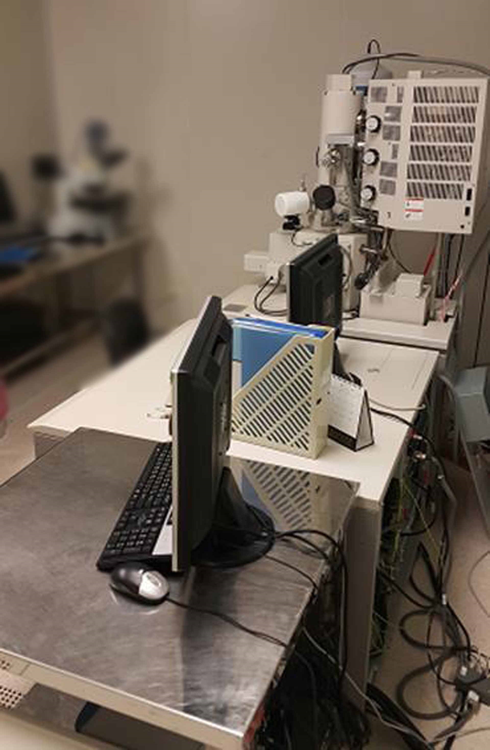
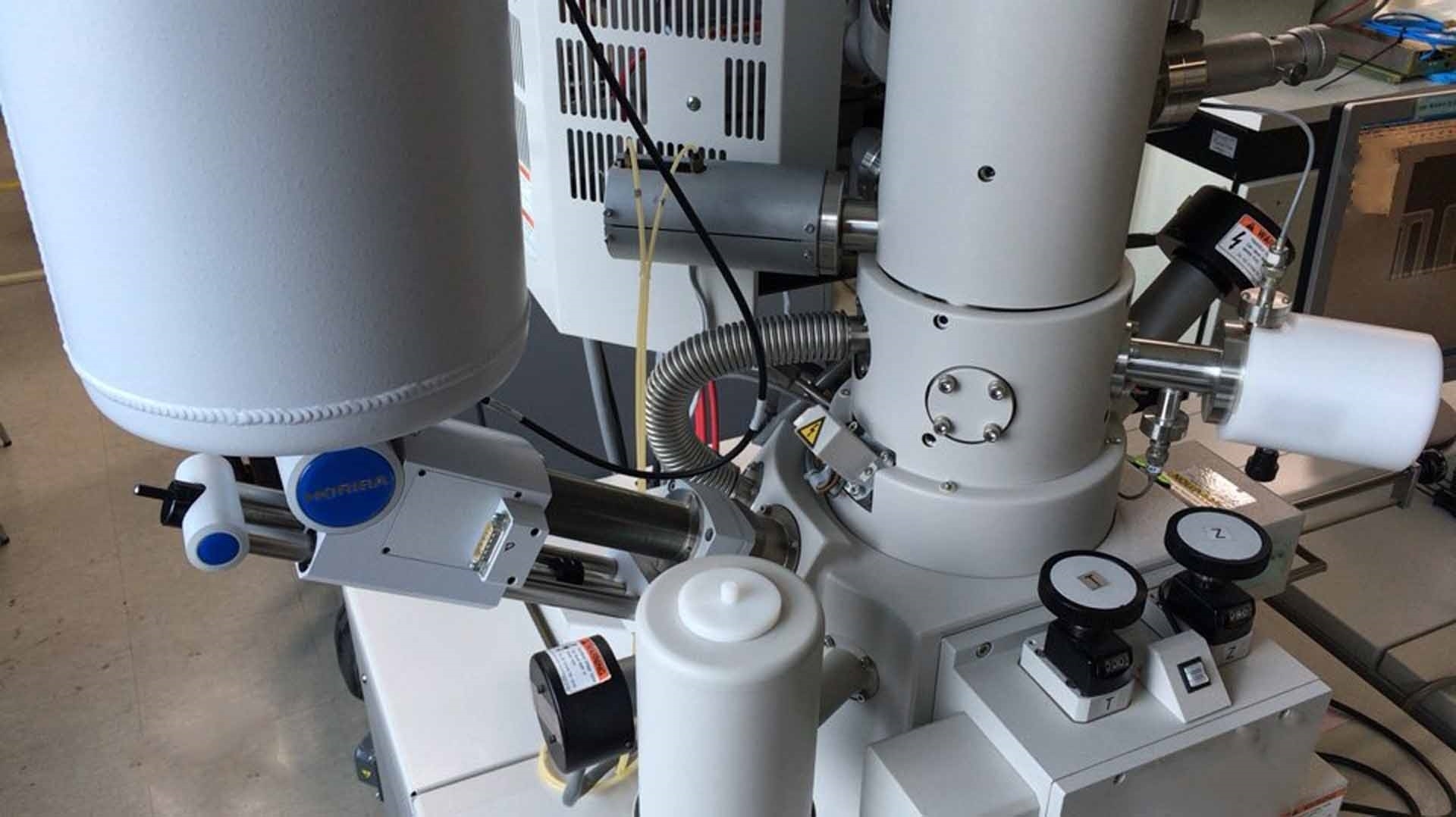
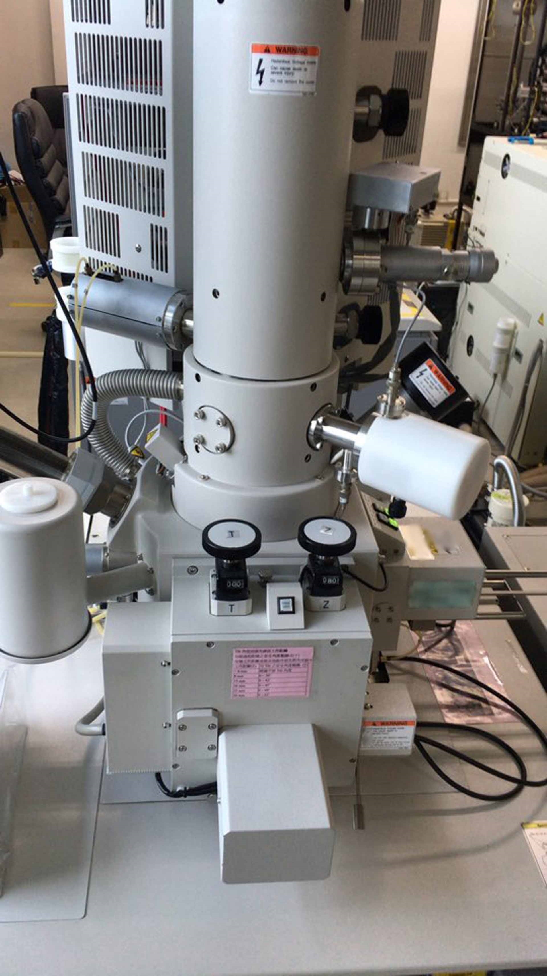
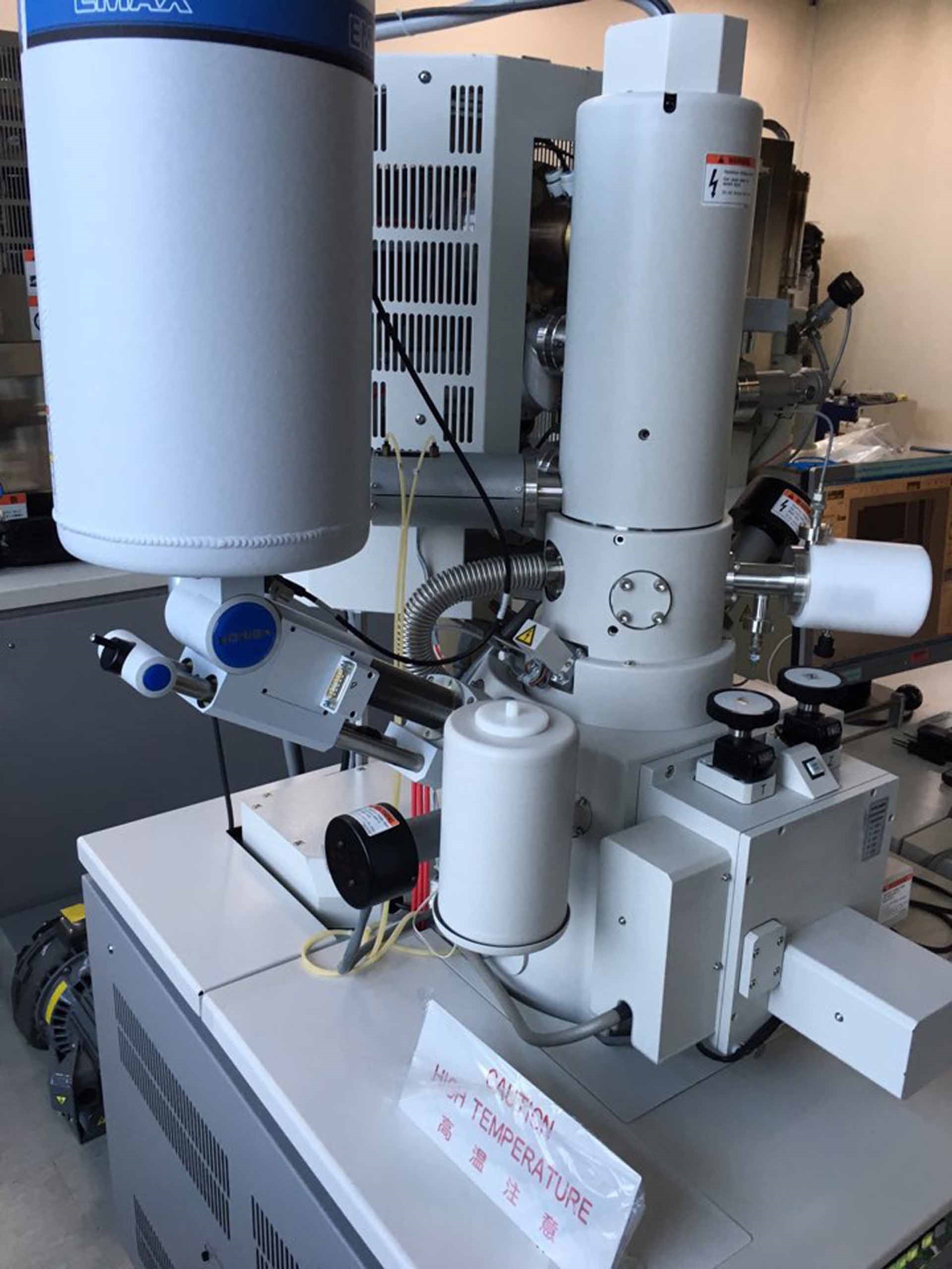
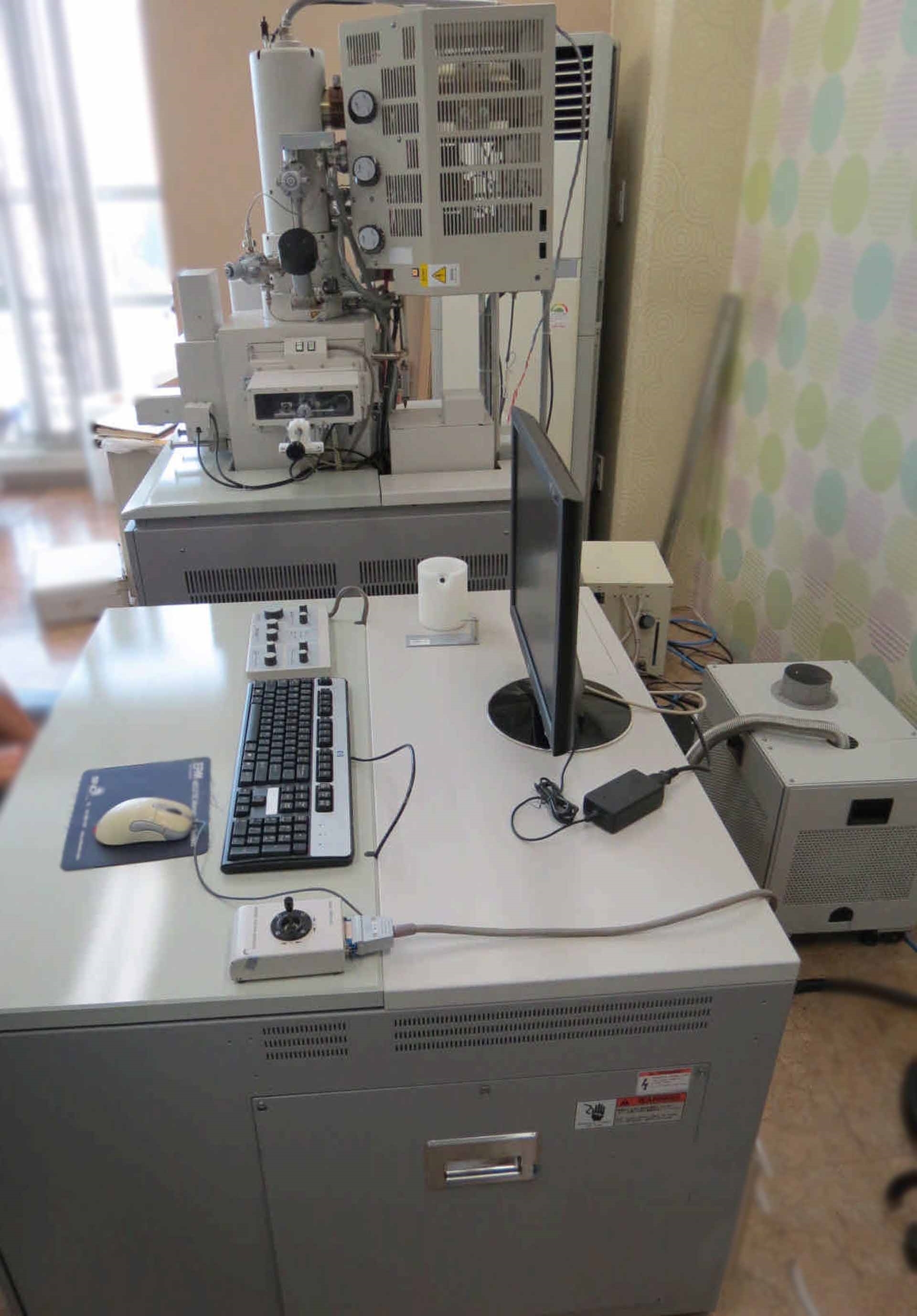
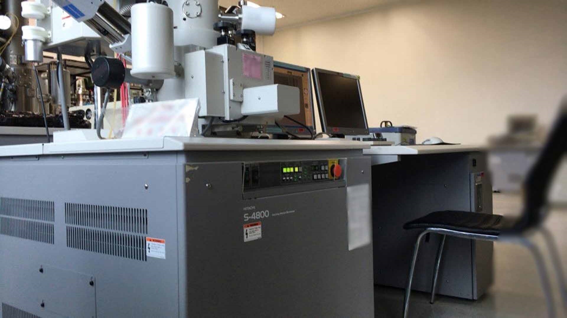
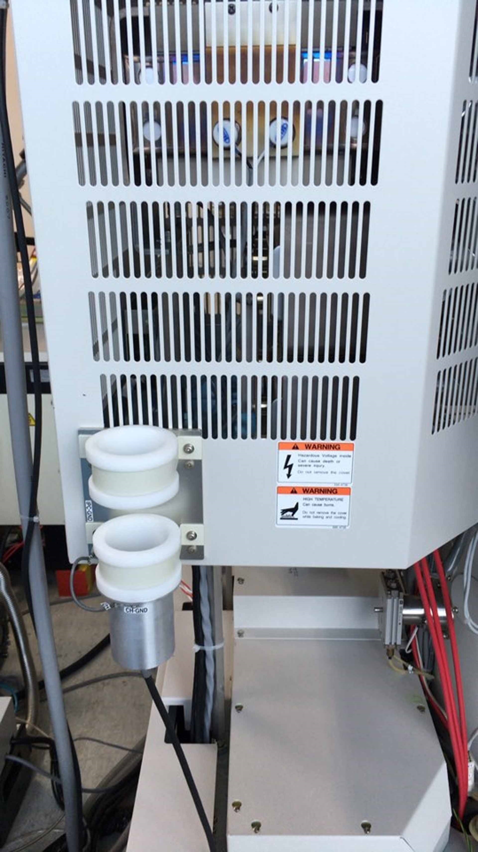
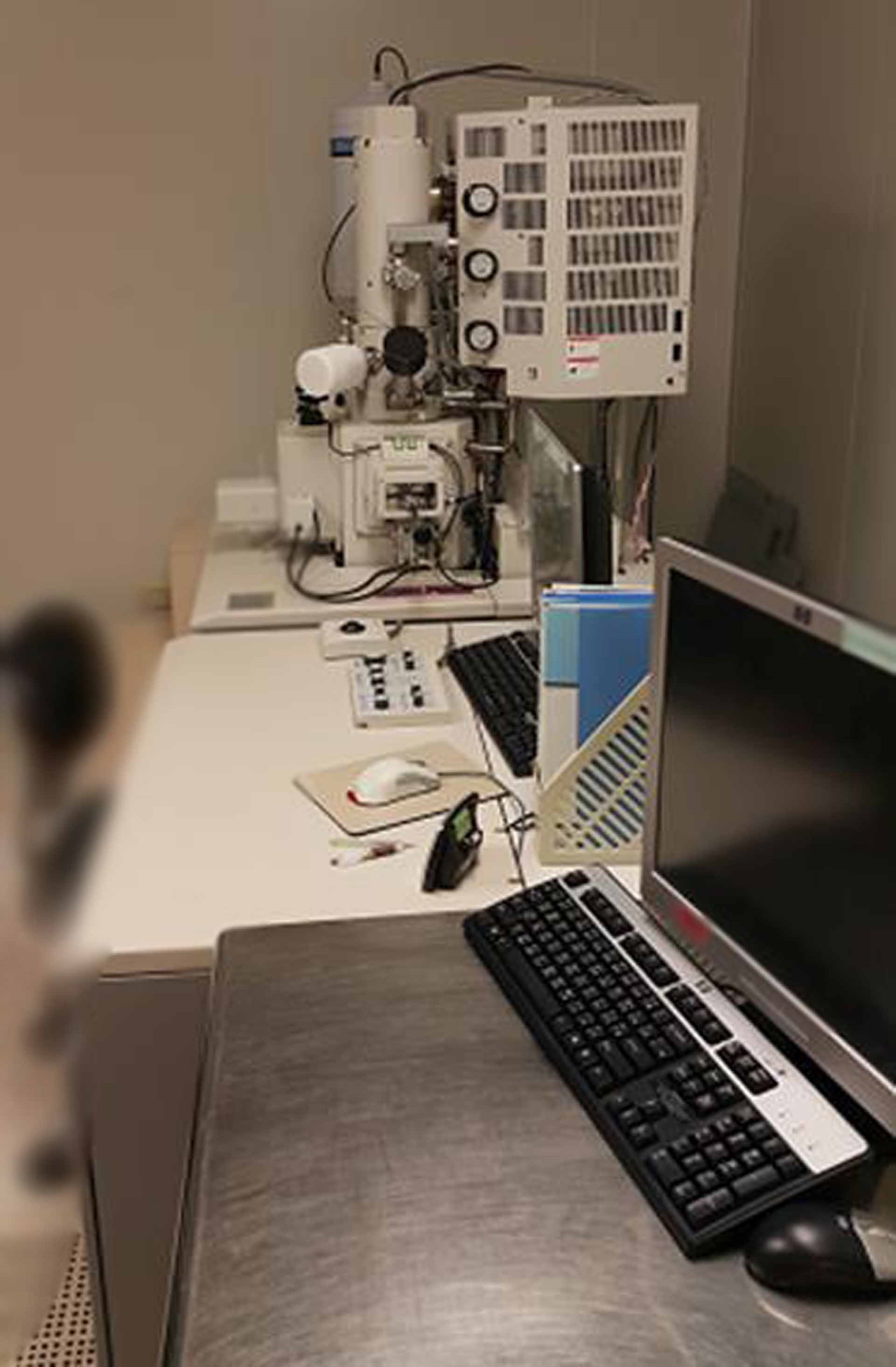
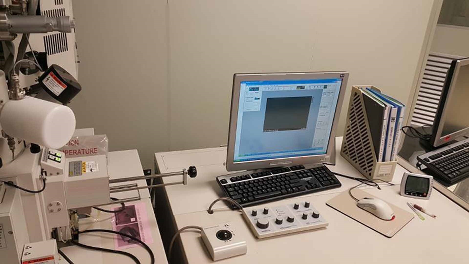
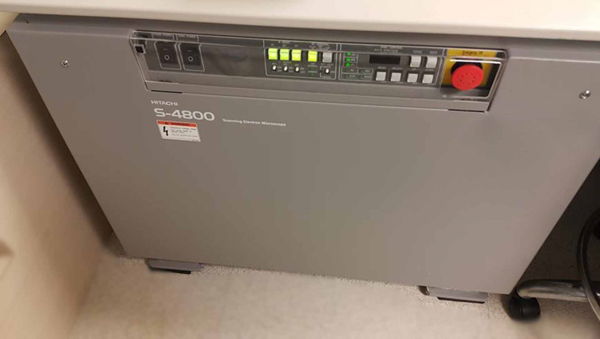
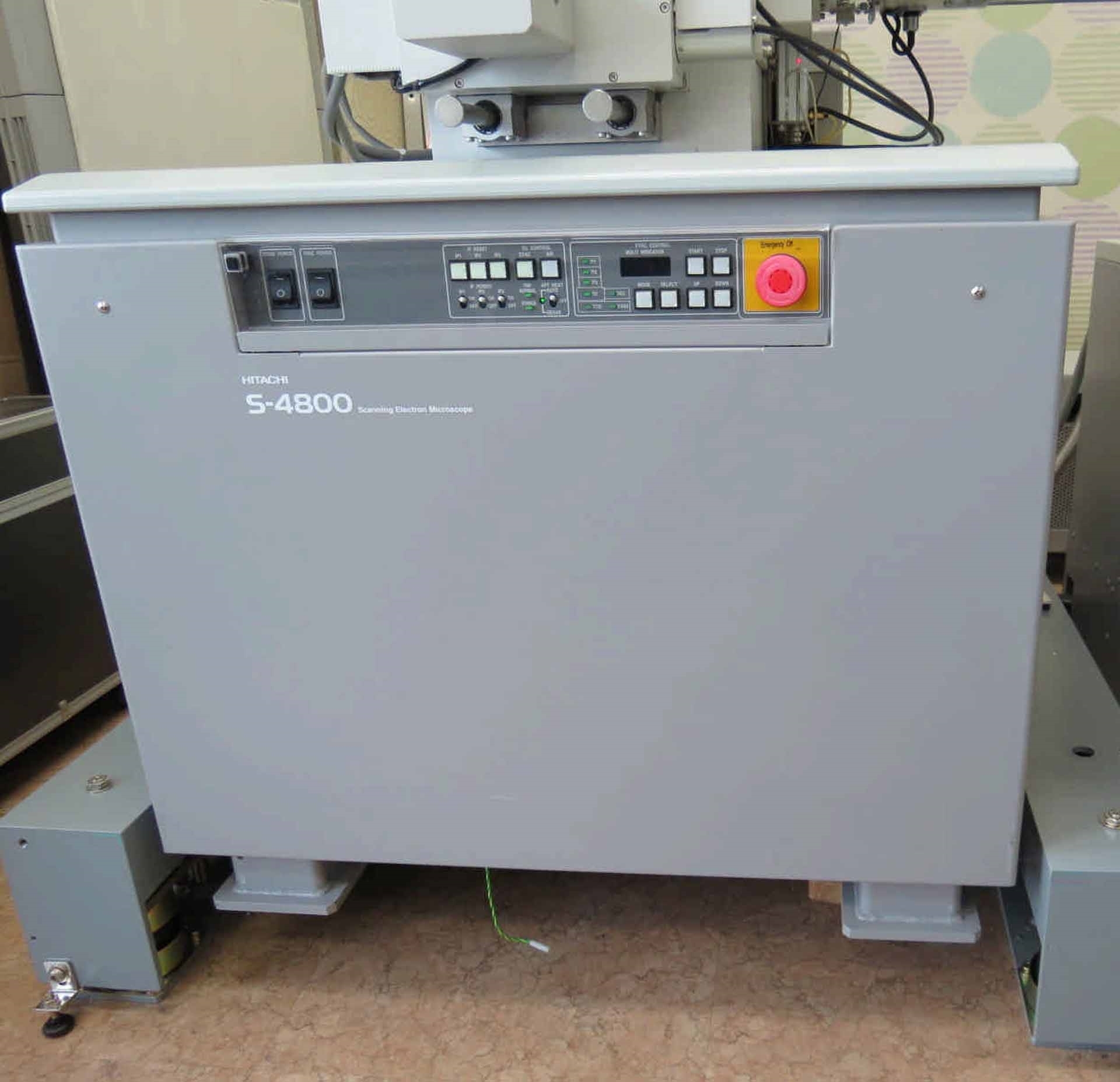
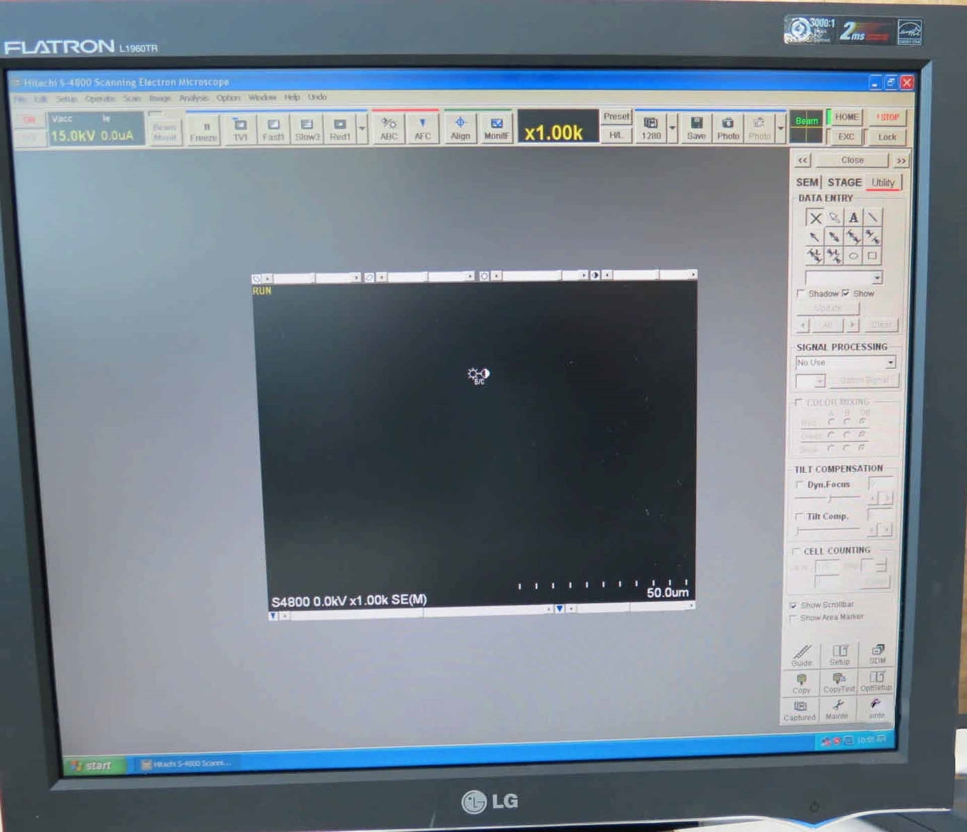
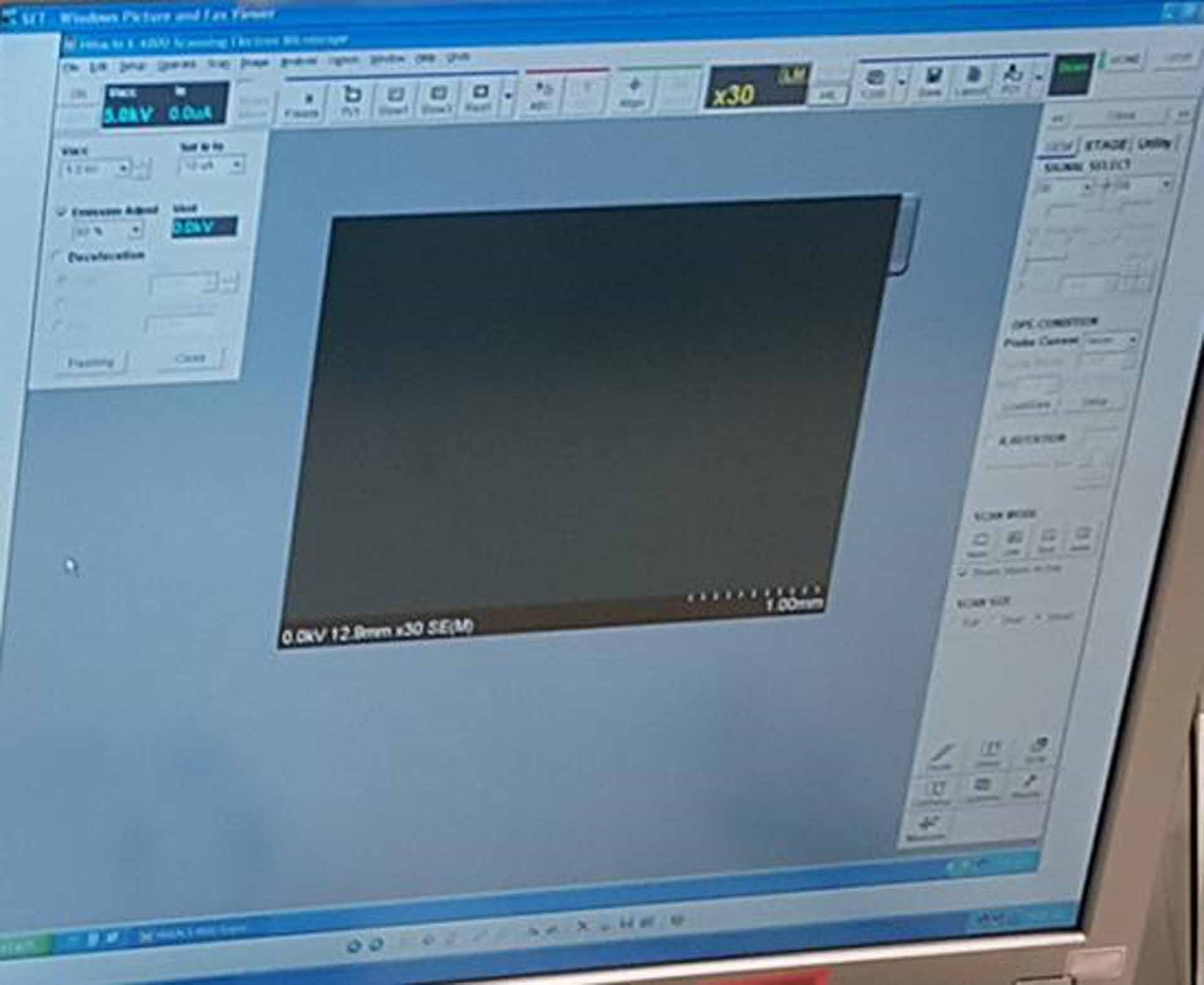
Field Emission Scanning Electron Microscope (FE-SEM)
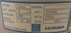
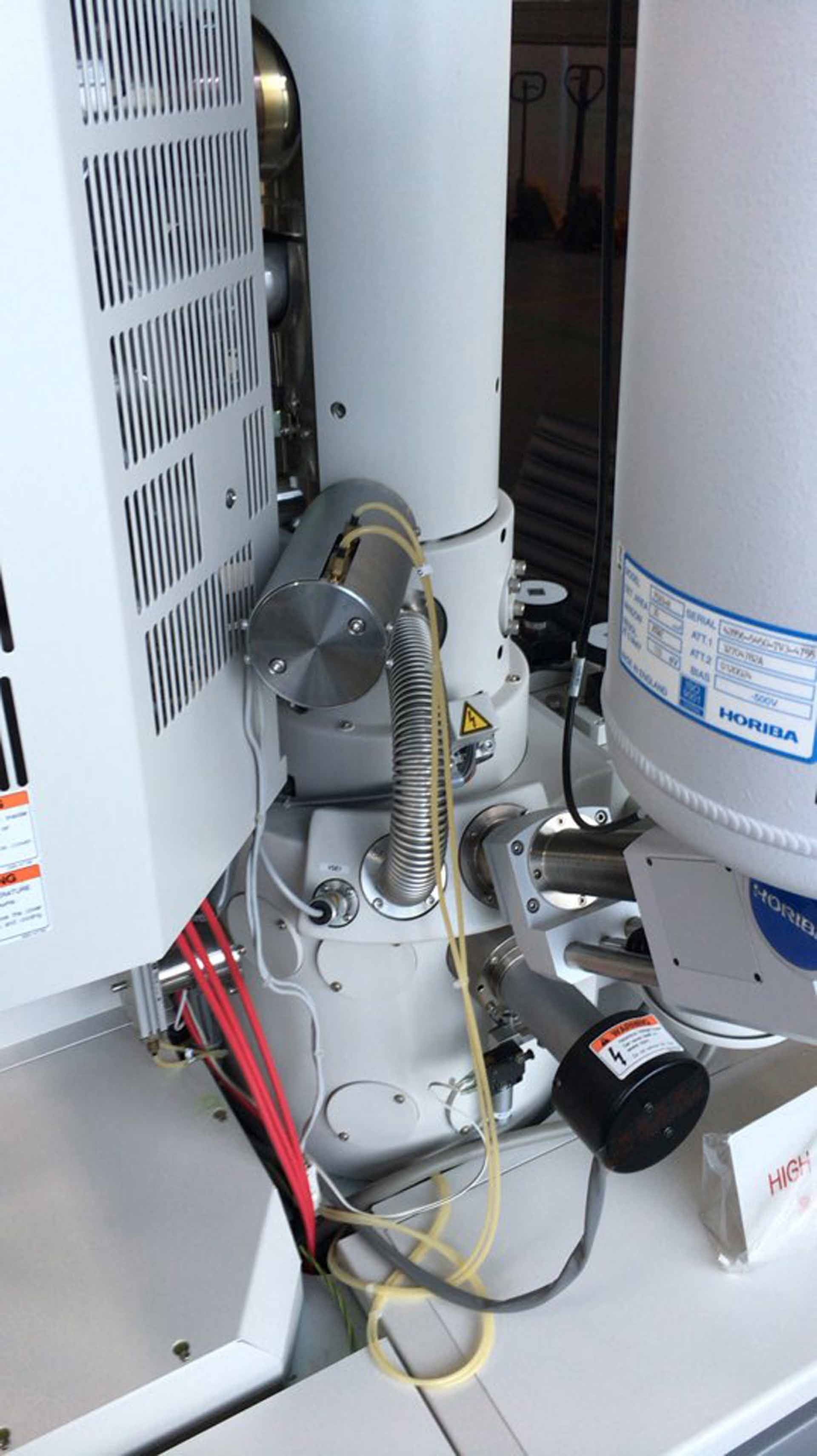
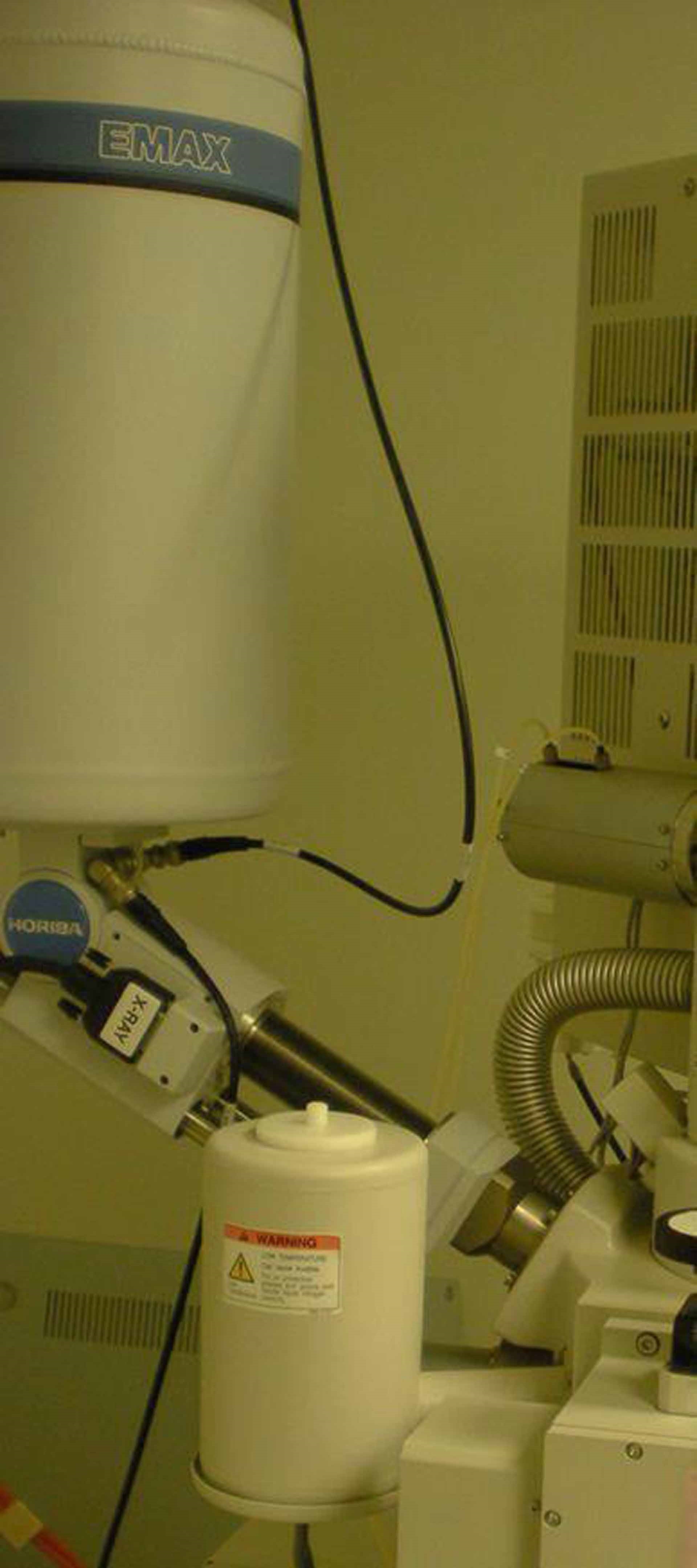
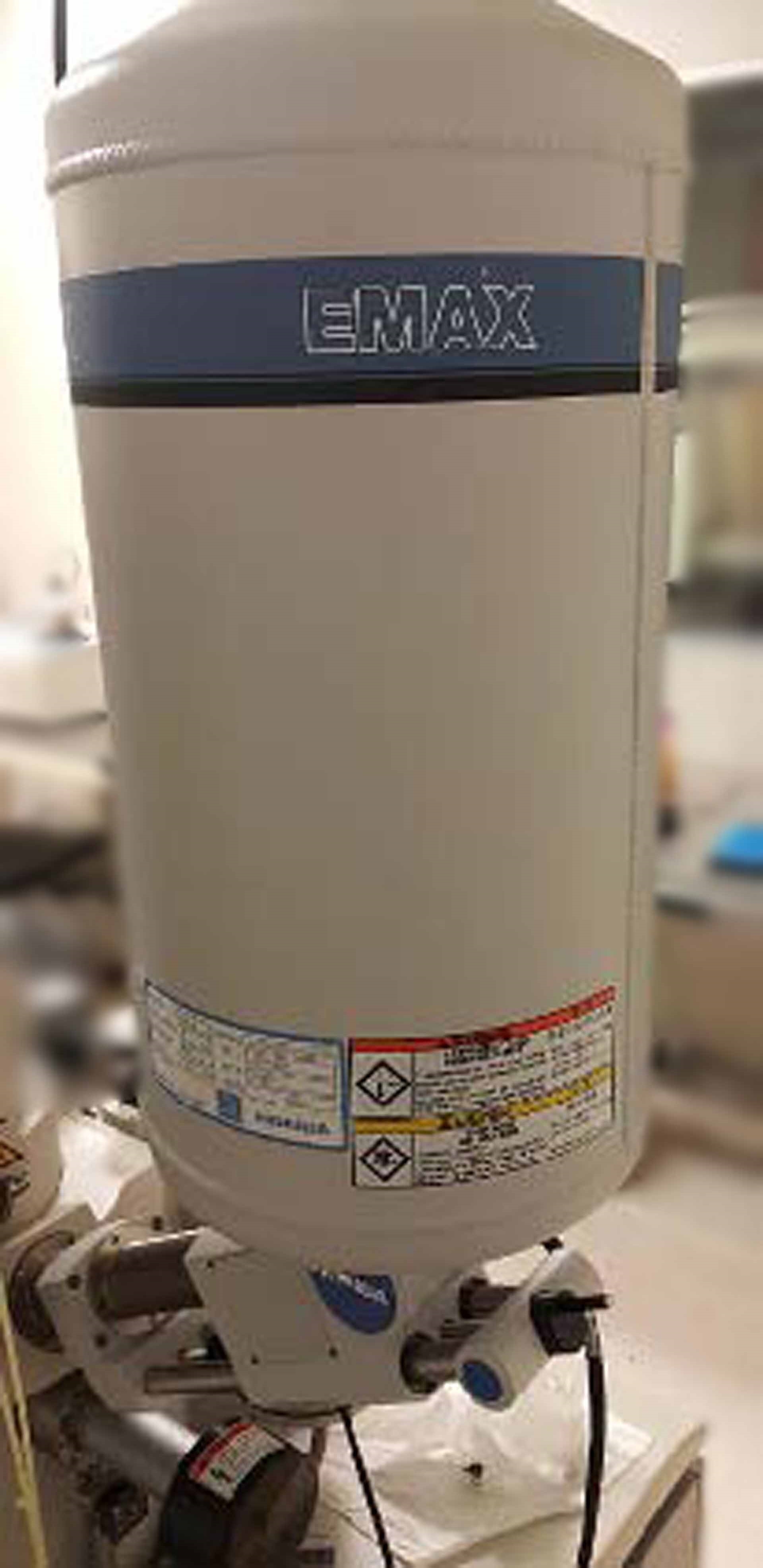
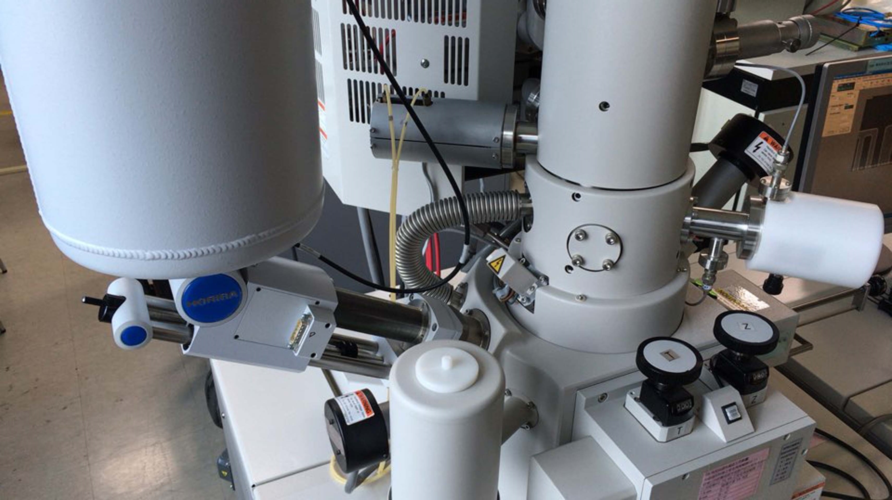
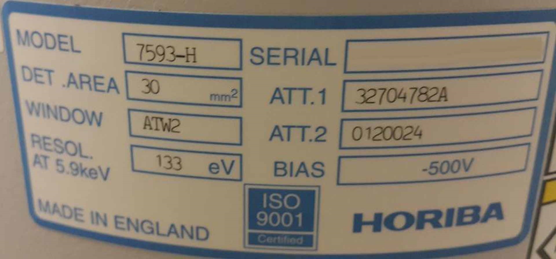
With HORIBA EDX system
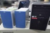
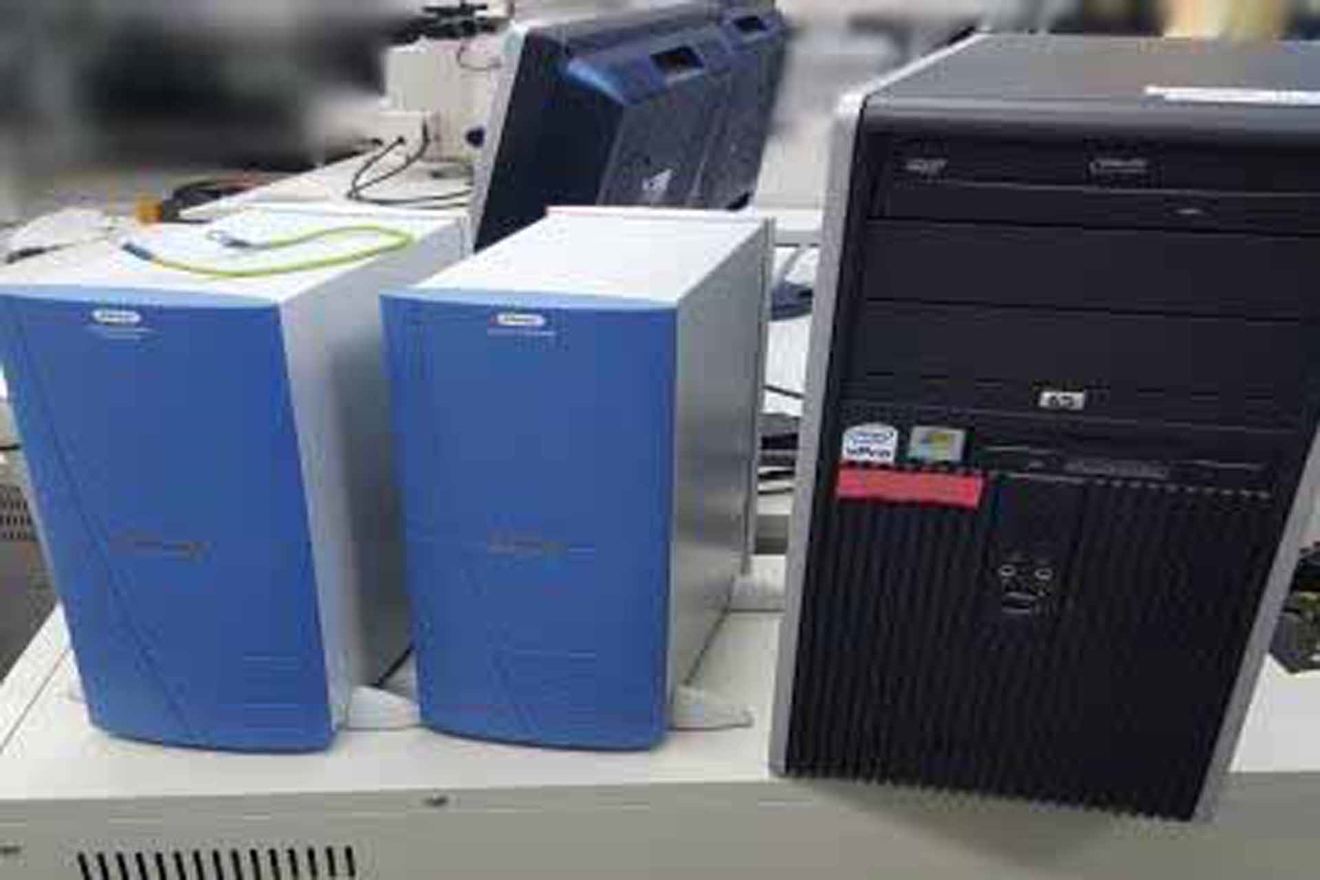
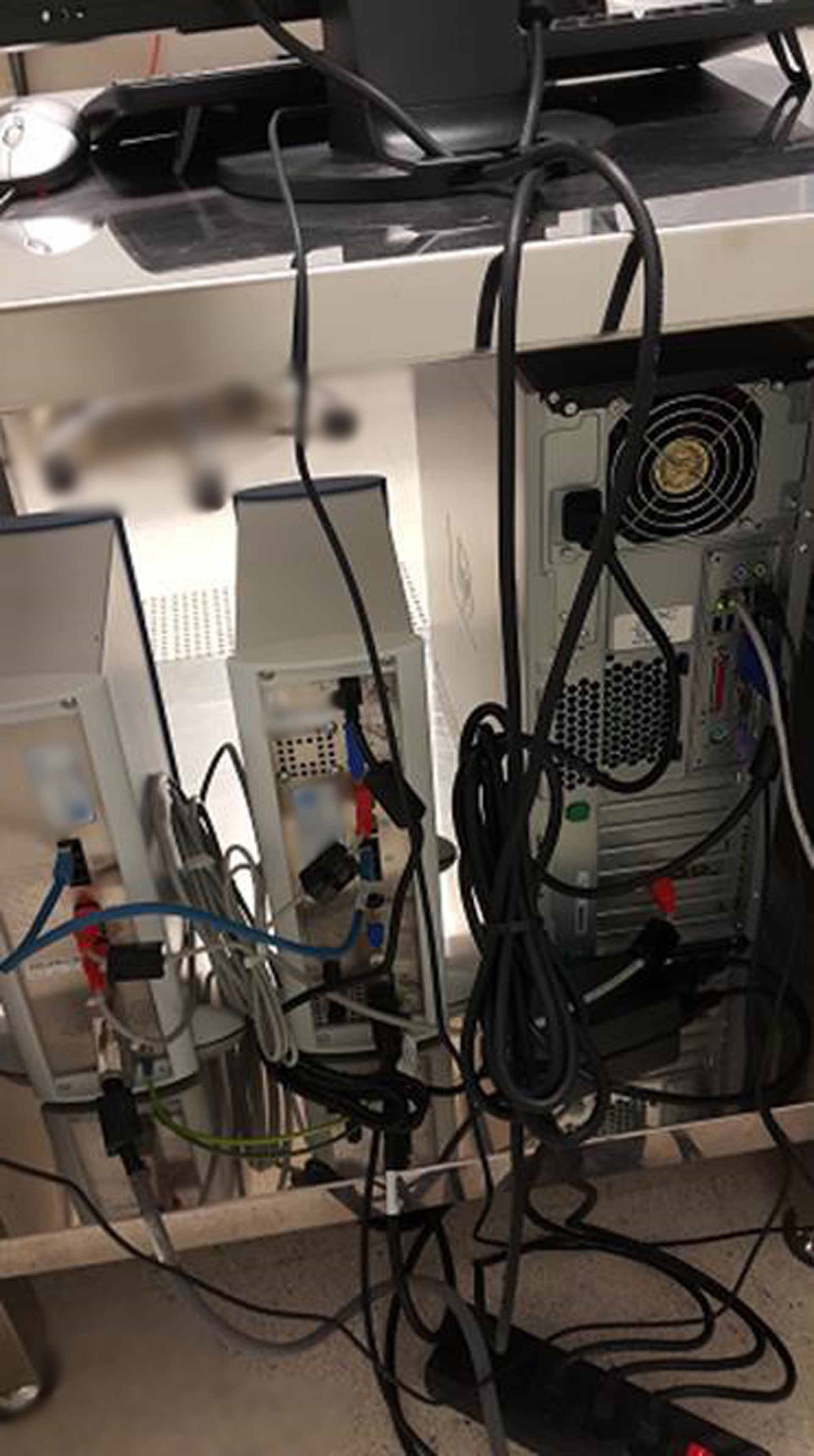
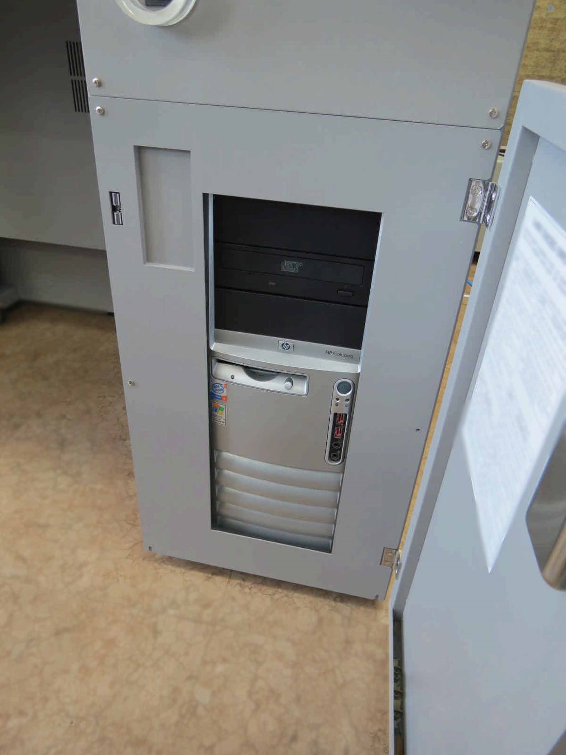
Workstation: HP DC7100MT
Declaration mode: 2.0 nm at 1 kV, WD: 1.5 mm, Normal mode
Stigmator: Octopole electromagnetic
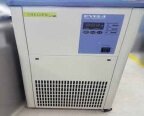
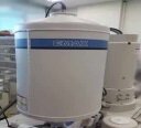
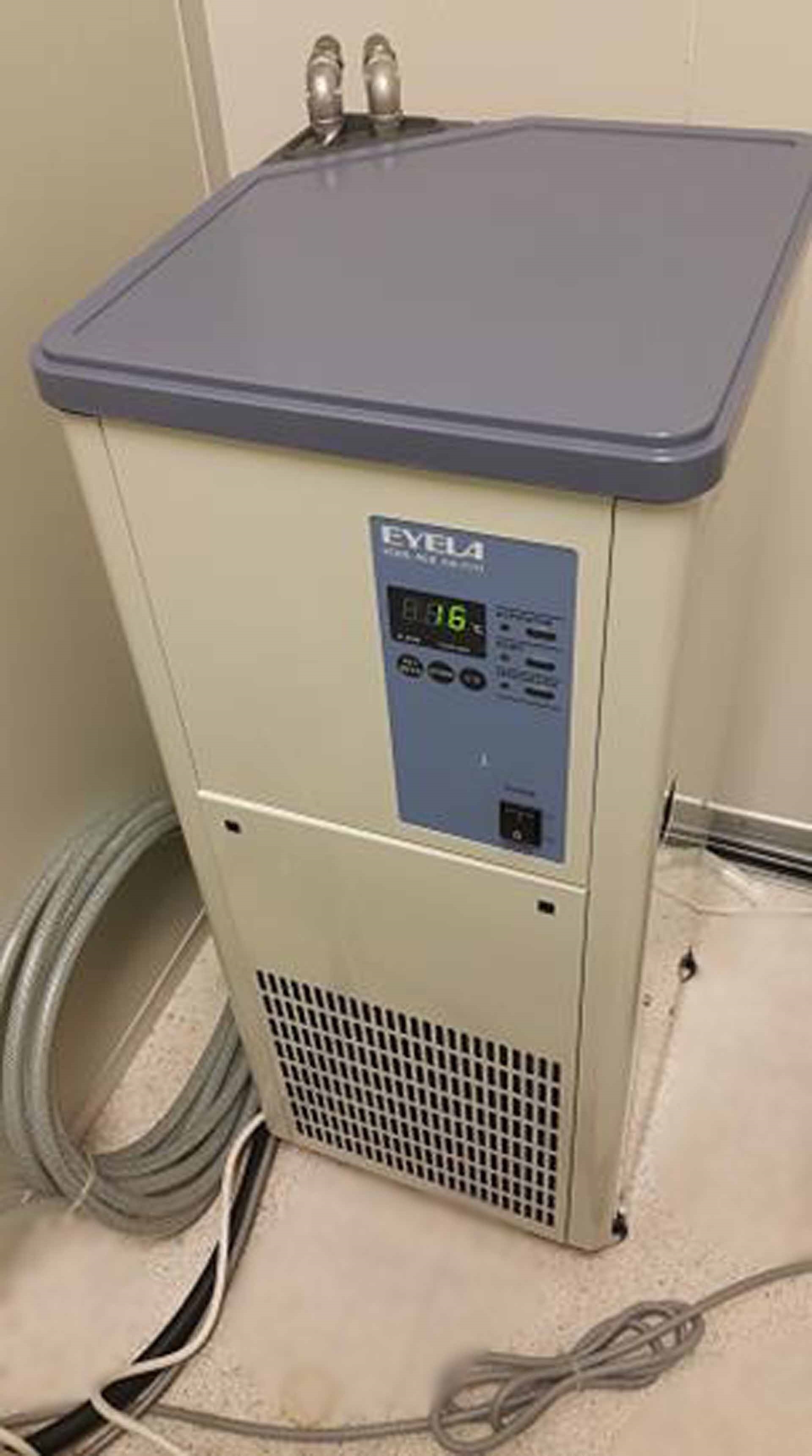
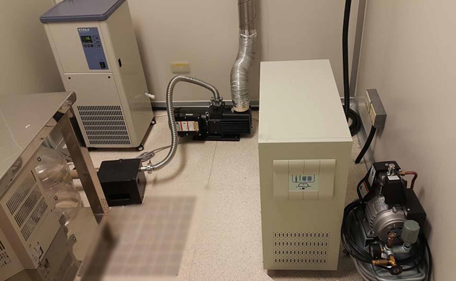
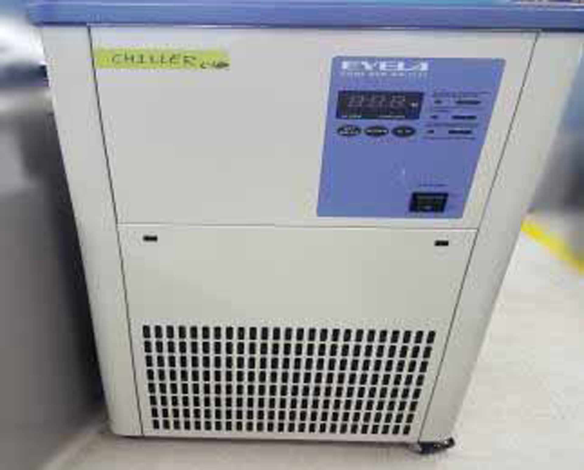
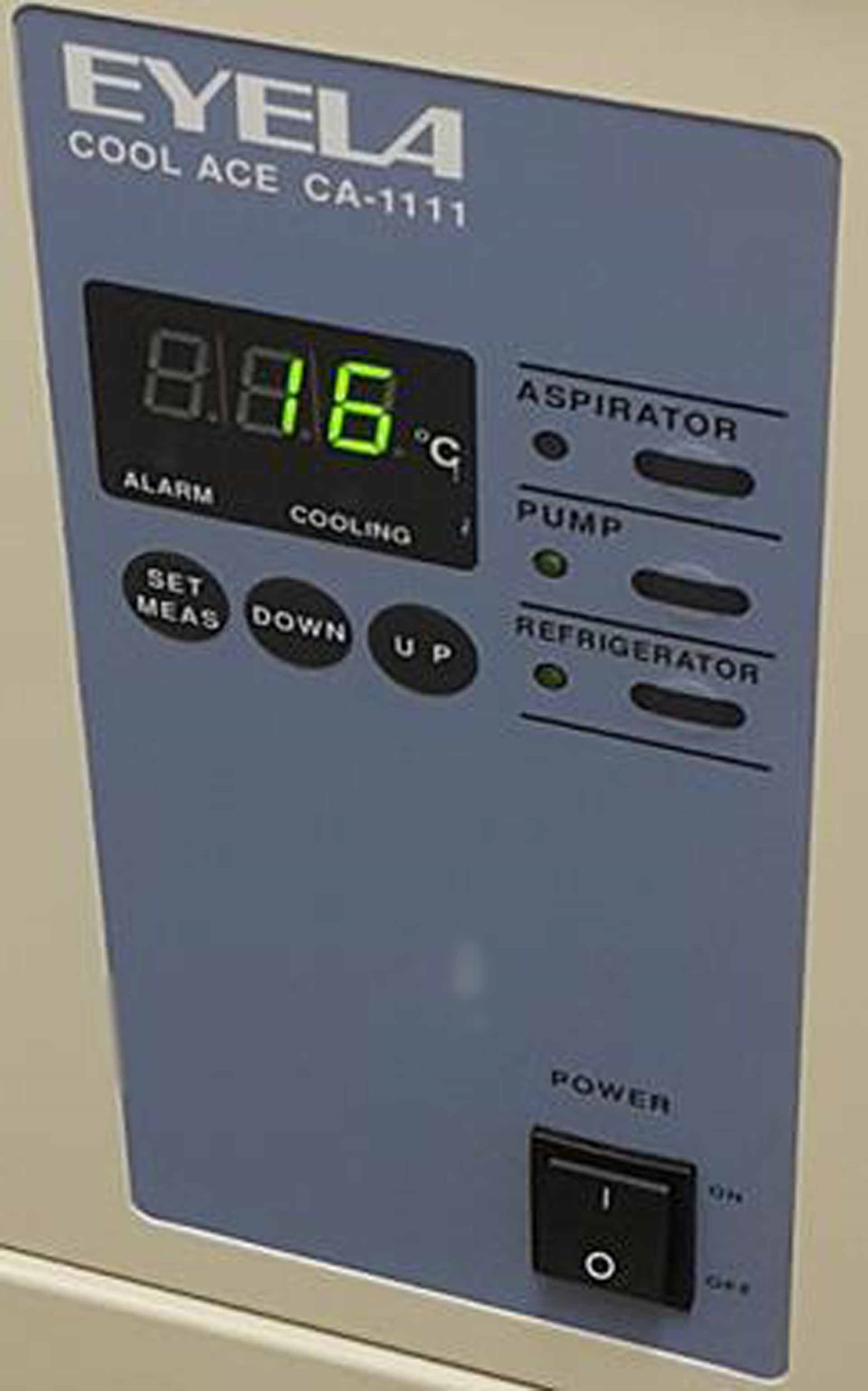
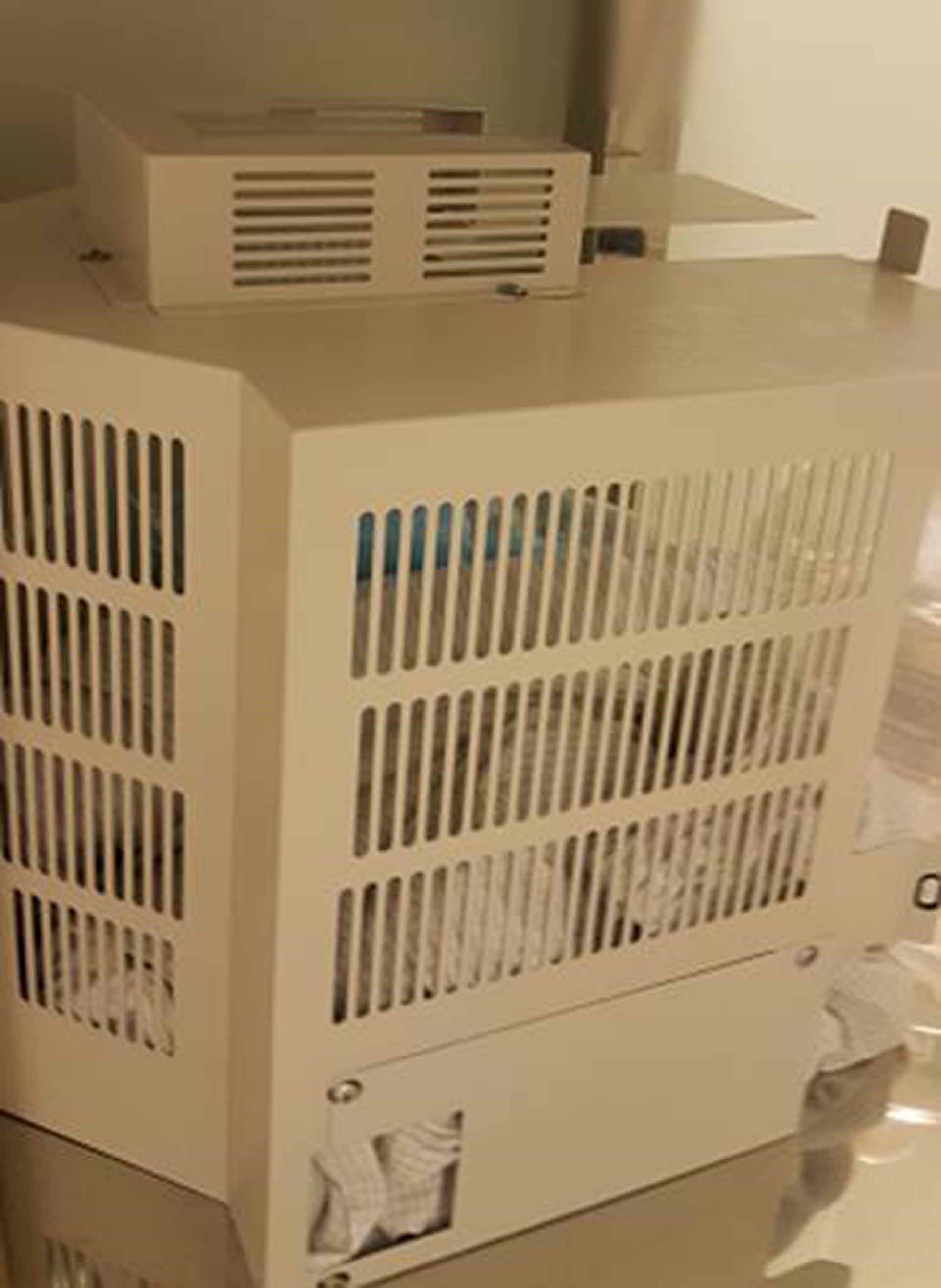
EYELA Cool Ace CA-1111 Chiller
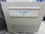
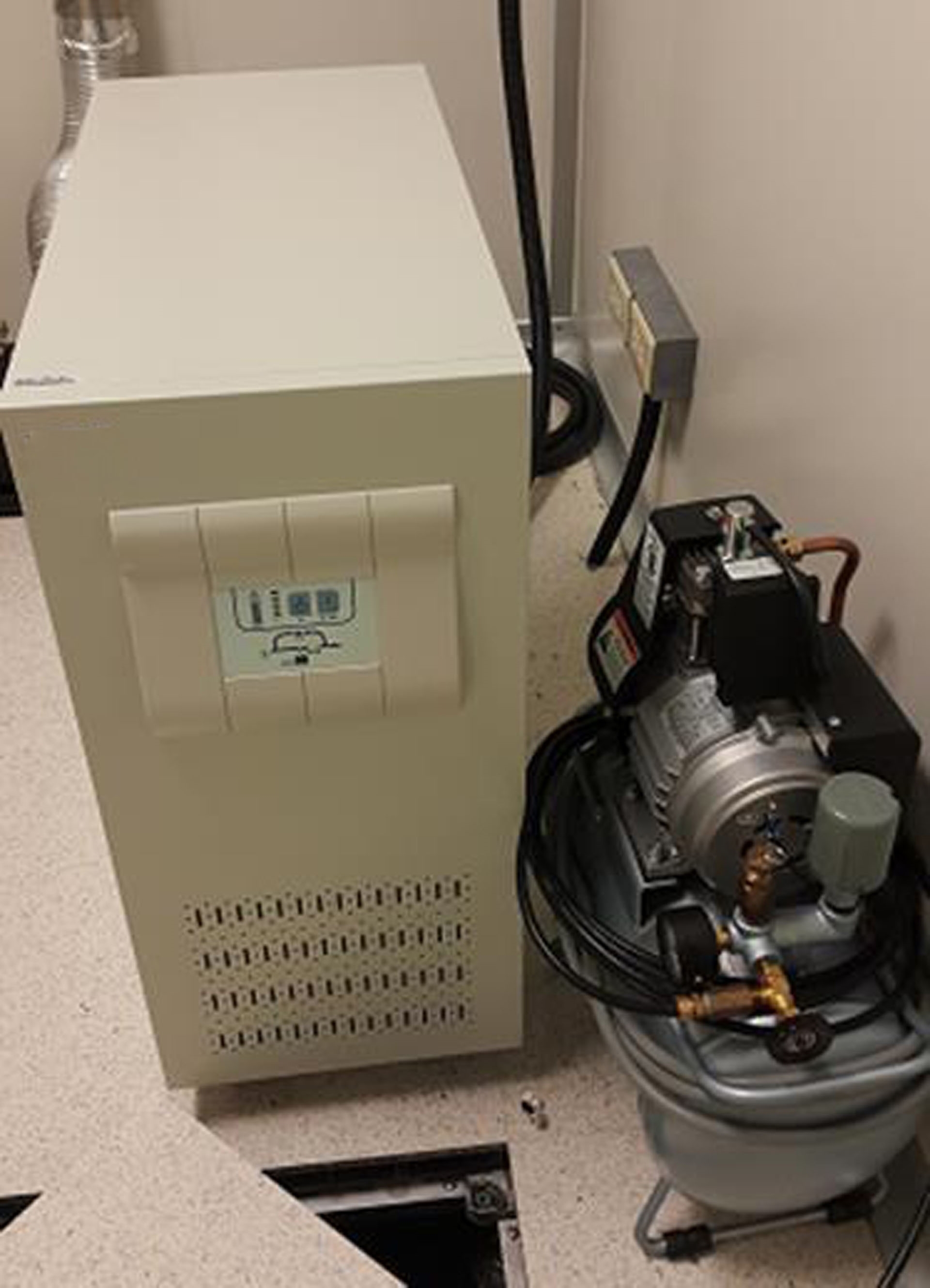
UPS
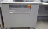
HITACHI Air compressor
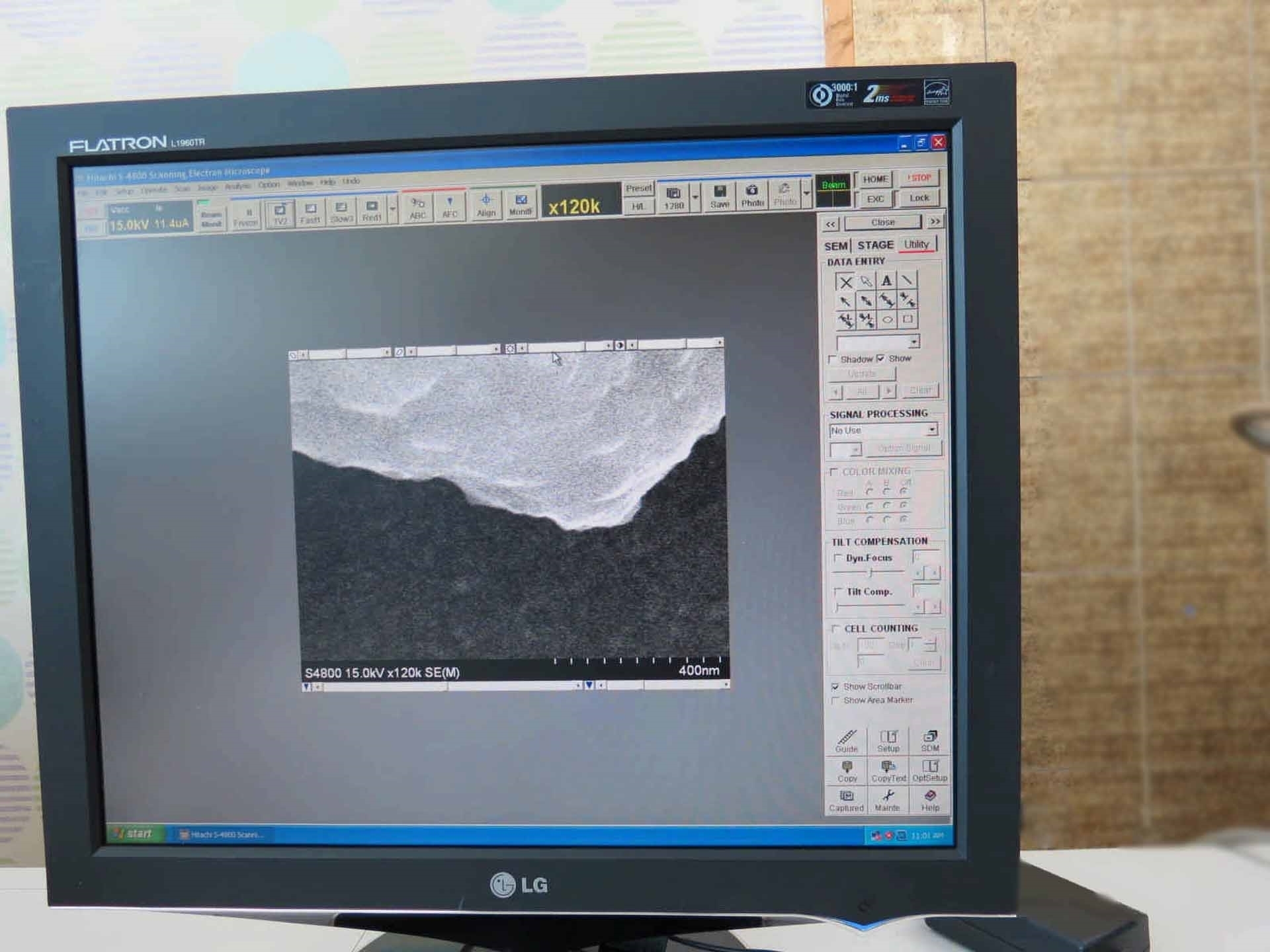
Operating system: Windows XP
Resolution:
Accelerating voltage: 15 kV
Working distance: 4 mm to 1.0 nm (220,000x)
Accelerating voltage: 1 kV
Working distance: 1.5 mm to 2.0 nm (120,000x)
Magnification:
High magnification mode: 100x to 800,000x
Low magnification mode: 20x to 2,000x
Electron optics:
Electron gun: Cold cathode field emission type
Extracting voltage (Vext): 0 to 6.5 kV
Accelerating voltage (Vacc): 0.5 to 30 kV (in 100 V steps)
Lens: 3-Stage electromagnetic lens, reduction type
Objective lens aperture:
Movable aperture (4 Openings selectable / Alignable outside column)
Self-cleaning thin aperture
Astigmatism correction coil: Electromagnetic type (Stigmator)
Scanning coil: 2-Stage electromagnetic electron optics
Specimen stage:
X-Traverse: 0 to 50 mm (Continuous)
Y-Traverse: 0 to 50 mm (Continuous)
Z-Traverse: 1.5 to 30.0 mm (Continuous)
Tilt: -5° to +70°
Rotation: 360° (Continuous)
Specimen size: Max 100 mm (Diameter)
(Airlock type specimen exchange)
Display unit:
Display type: Flicker-free image on PC monitor (Full scanning speeds)
Viewing monitor: Type 18.1 LCD
Option: Type 21 Color CRT (1280 x 1024 pixels)
Photo CRT (Option): Ultra-high resolution type
(Effective field of view 120 x 90 mm)
Full screen: 1280 x 960 Pixels
Reduced area:
640 x 480 Pixels
320 x 240 Pixels
Dual Image: 640 x 480 Pixels
Scanning modes:
Normal scan
Reduced area scan
Line scan
Spot analysis
Average concentration analysis
Split / Dual magnification
Scanning speeds:
TV (640 x 480 pixel display: 25 / 30 frames/s)
Fast (Full screen display: 6.25 / 7.5 frames/s)
Slow:
(Full screen display: 1 / 0.9 , 4 / 3.3 , 20 / 16, 40 / 32 ,80 / 64 Frames/s)
(640 x 480 pixels display: 0.5 / 0.4 , 2 / 1.7 , 10 / 8 , 20 / 16, 40 / 32 Frames/s)
Photograph: 2560 x 1920 Pixels
Display: 40 / 32, 80 / 64, 160 / 128, 320 / 256 Frames/s
Value: 50 Hz / 60 Hz
TV: NTSC or PAL Signal
Signal processing modes:
Automatic brightness control
Gamma control
Automatic focus
Automatic stigmator
Automatic data display:
Image number
Accelerating voltage
Magnification
Micron bar
Micron value
Data / Time
Data entry: Alphanumeric characters, number, and marks
Electrical image shift: 12 m (WD: 8 mm)
Evacuation system:
System type: Fully automatic pneumatic-valve system
Ultimate vacuum levels: Specimen chamber: 7 x Pa
10-7 Pa in electron gun chamber
10-4 Pa in specimen chamber
Electron gun chamber:
IP1 1 x Pa or better
IP2 2 x Pa or better
IP3 7 x Pa or better
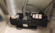
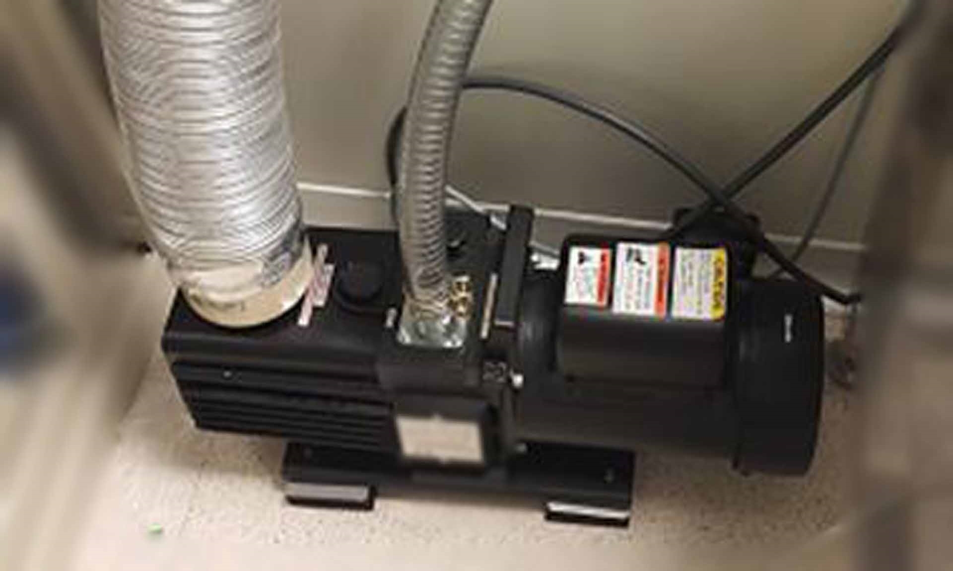
Vacuum pumps: ULVAC GLD-136
Electron optical system: (3) Ion pumps
Specimen chamber: Turbo molecular pump
Oil rotary pump
Protection devices:
Warning devices:
Power failure
Cooling-water interruption
Inadequate vacuum
Secondary electron image resolution:
1.0 nm at 15 kV, WD: 4 mm
1.4 nm at 1 kV, WD: 1.5 mm
Sample chamber:
Size: Type I stage
Max sample size: 100 mm Diameter
Stage motion:
3 Axis motorized
X/Y: 0 - 50 mm
Signal selection:
SE Signal
X-Ray signal
AUX Signal
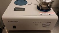
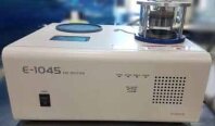
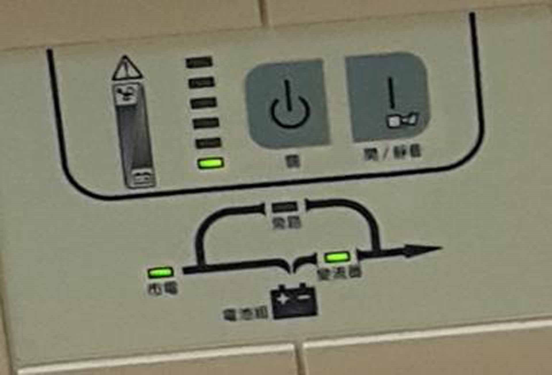
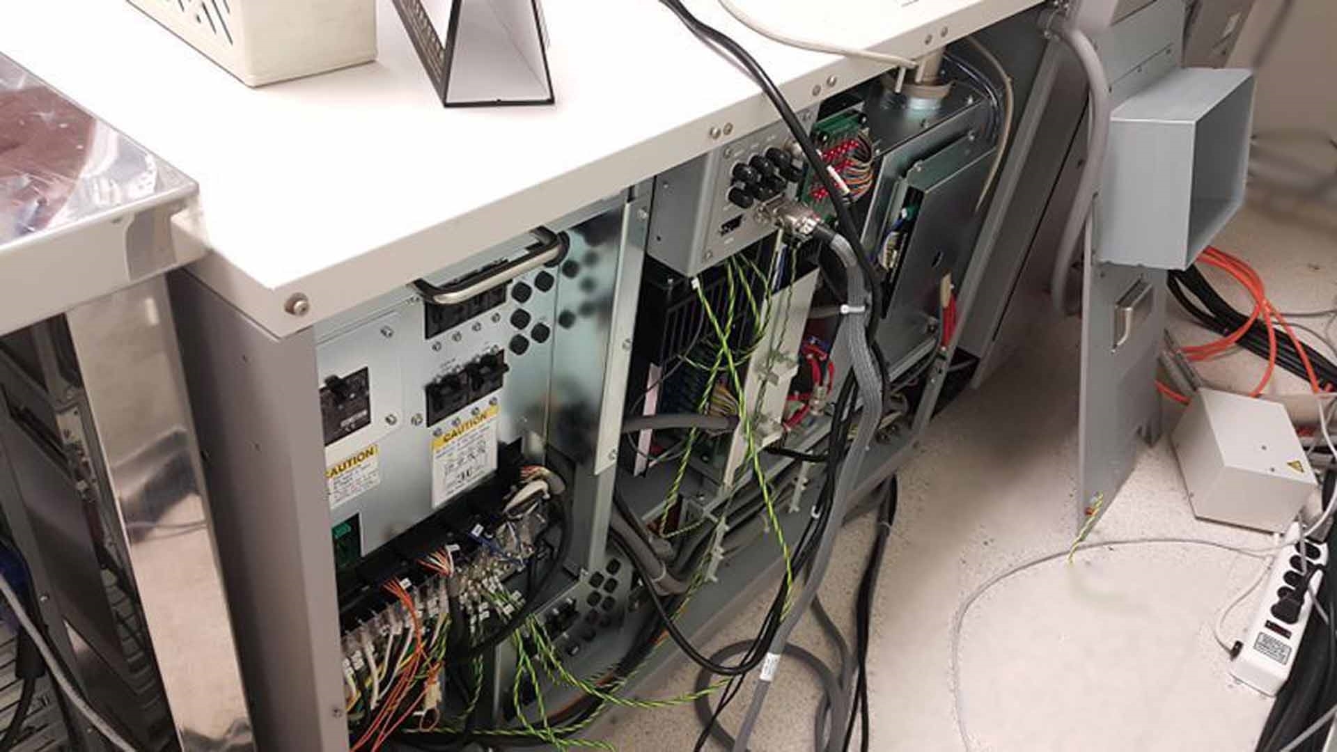
UPS Unit
Chiller
FE-SEM Rotary pump
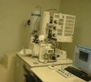
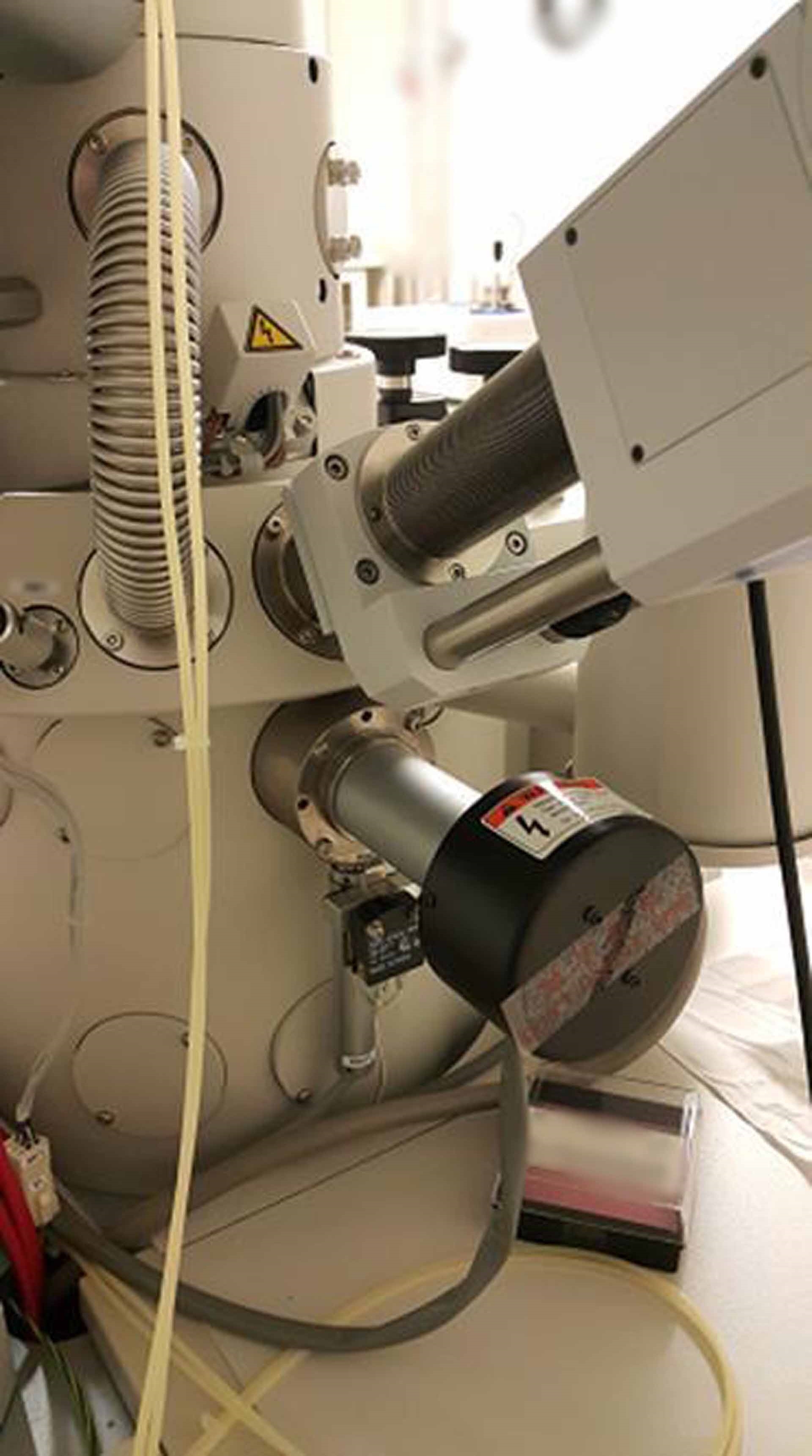
Main body:
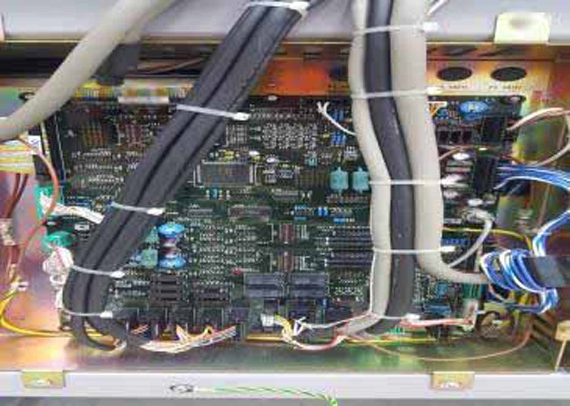
Stage controller (ROM PCB)
EVAC Controller (ROM PCB)
(3) Ion pumps
SE Detector
Multi-aperture
Gun head cap
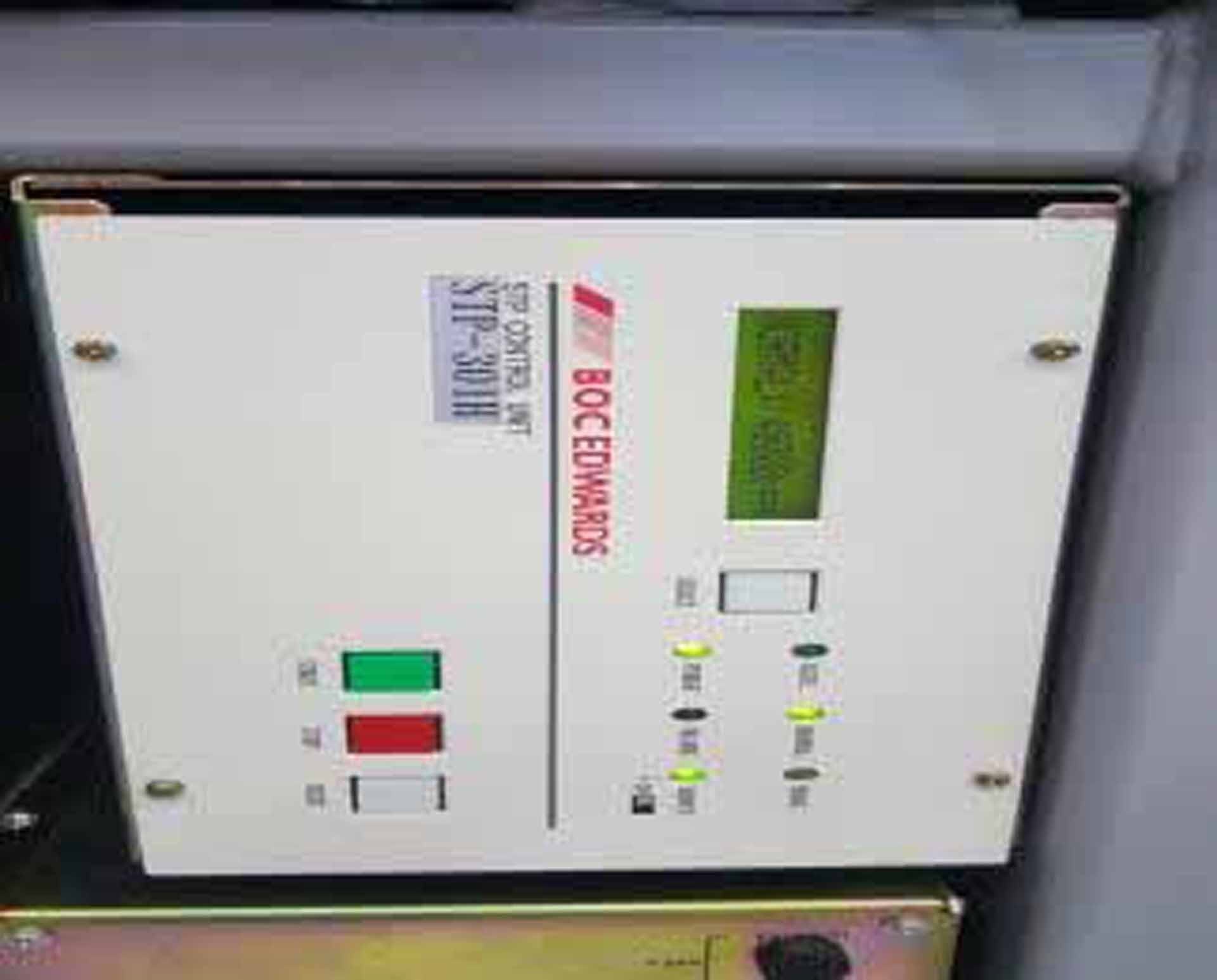
STP301H TMP Pump
STP301H TMP Controller
Solenoid valve assy
PC
Hard Disk Drive (HDD)
Operating system: Windows XP
HV Controller
Gun head unit
Case
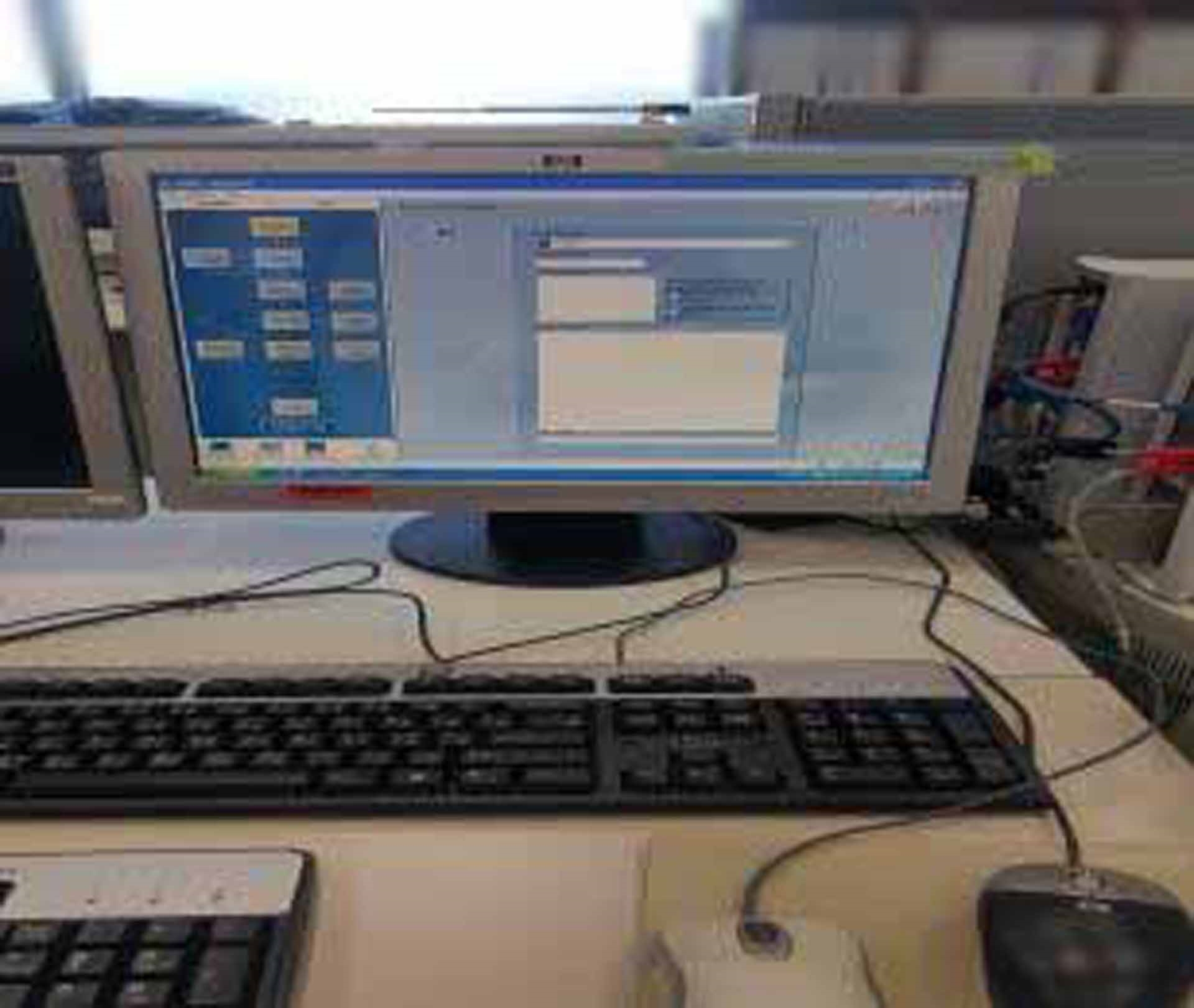
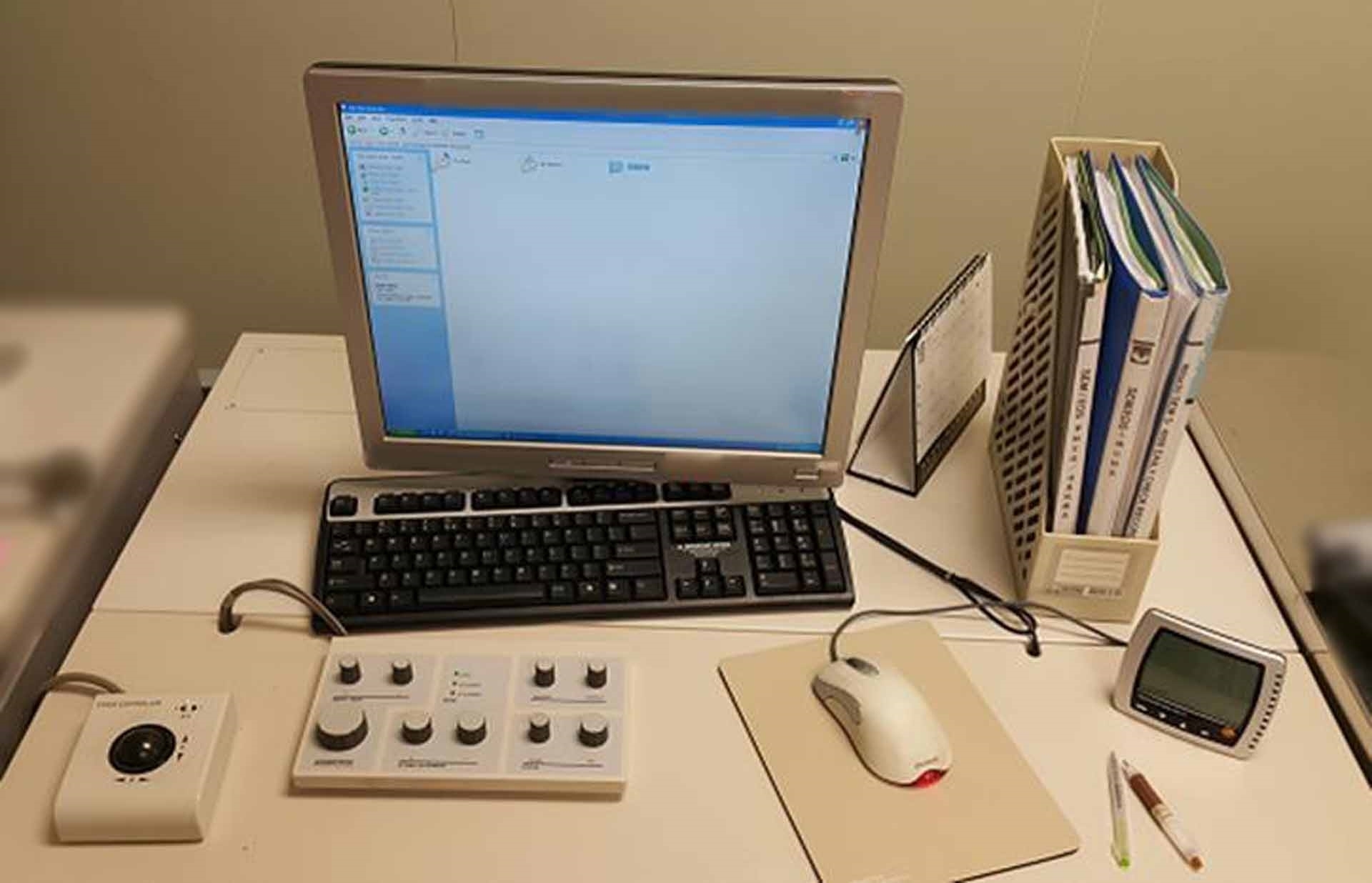
Operation unit:
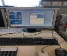
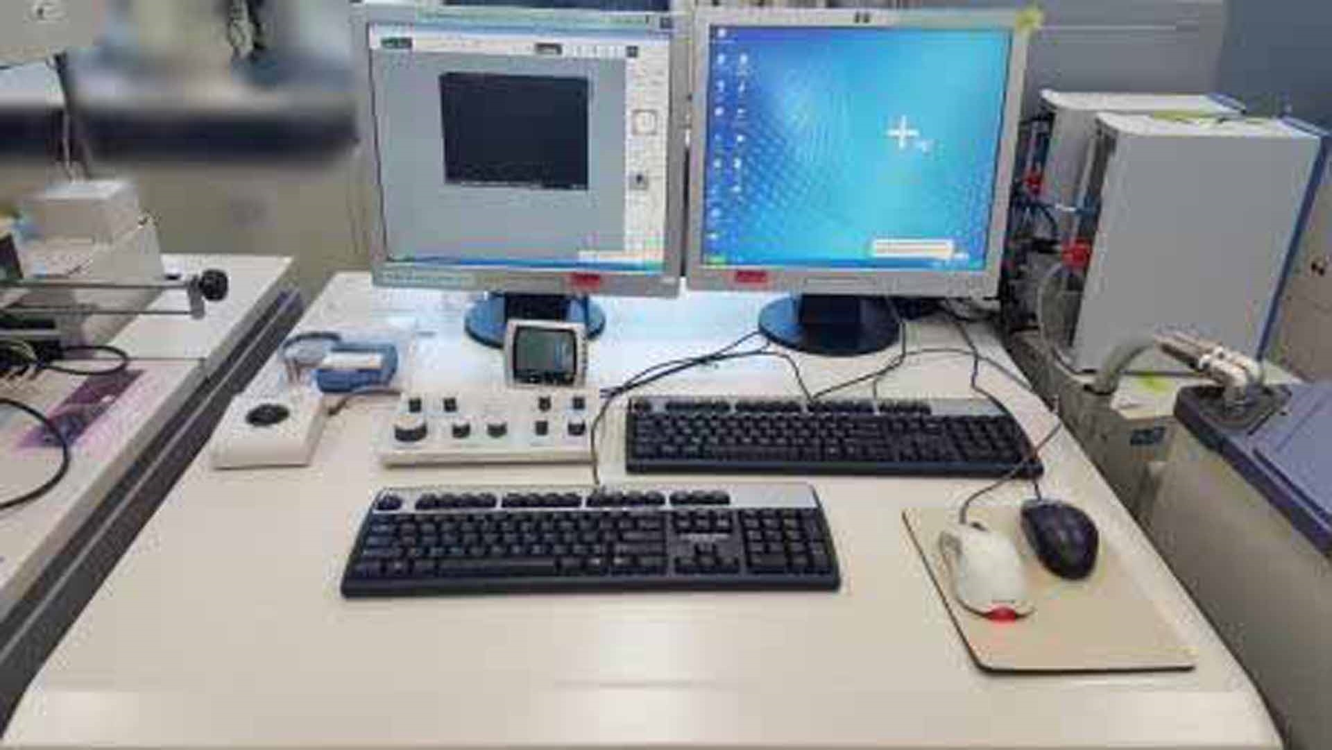
LCD Monitor
Keyboard
Mouse
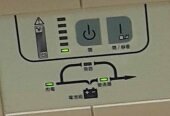
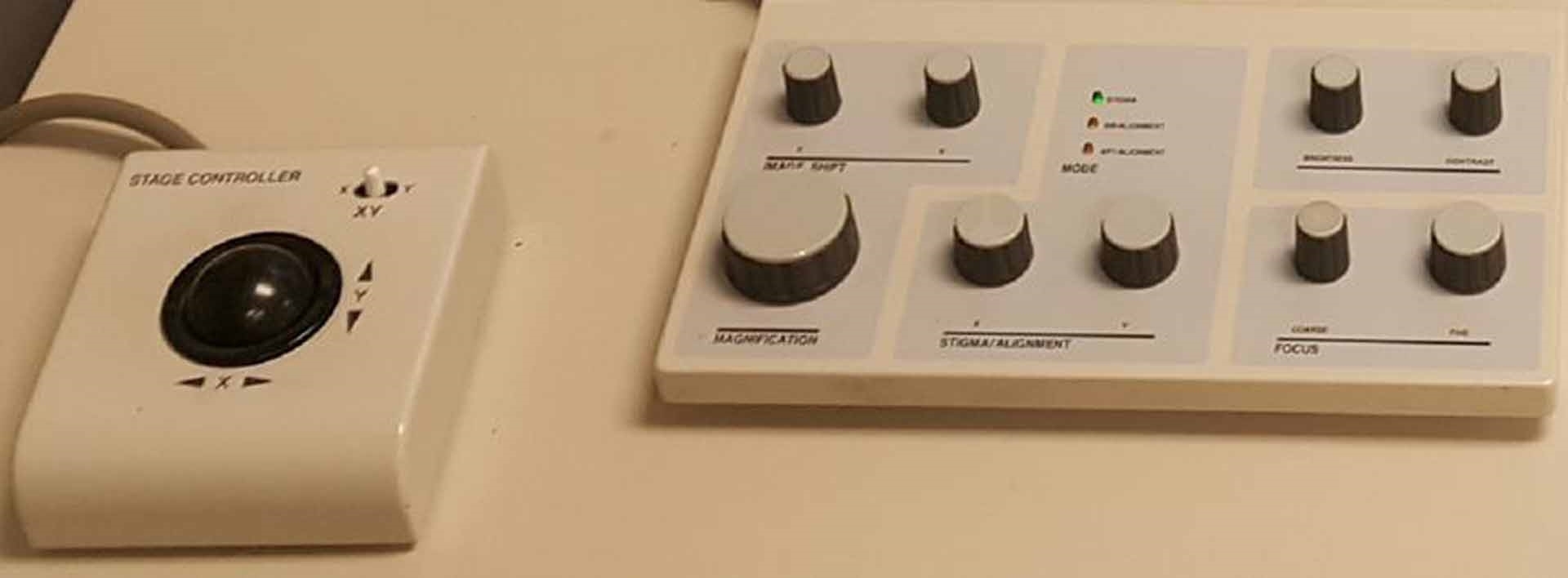
Operation panel
Stage control trackball
ETC:
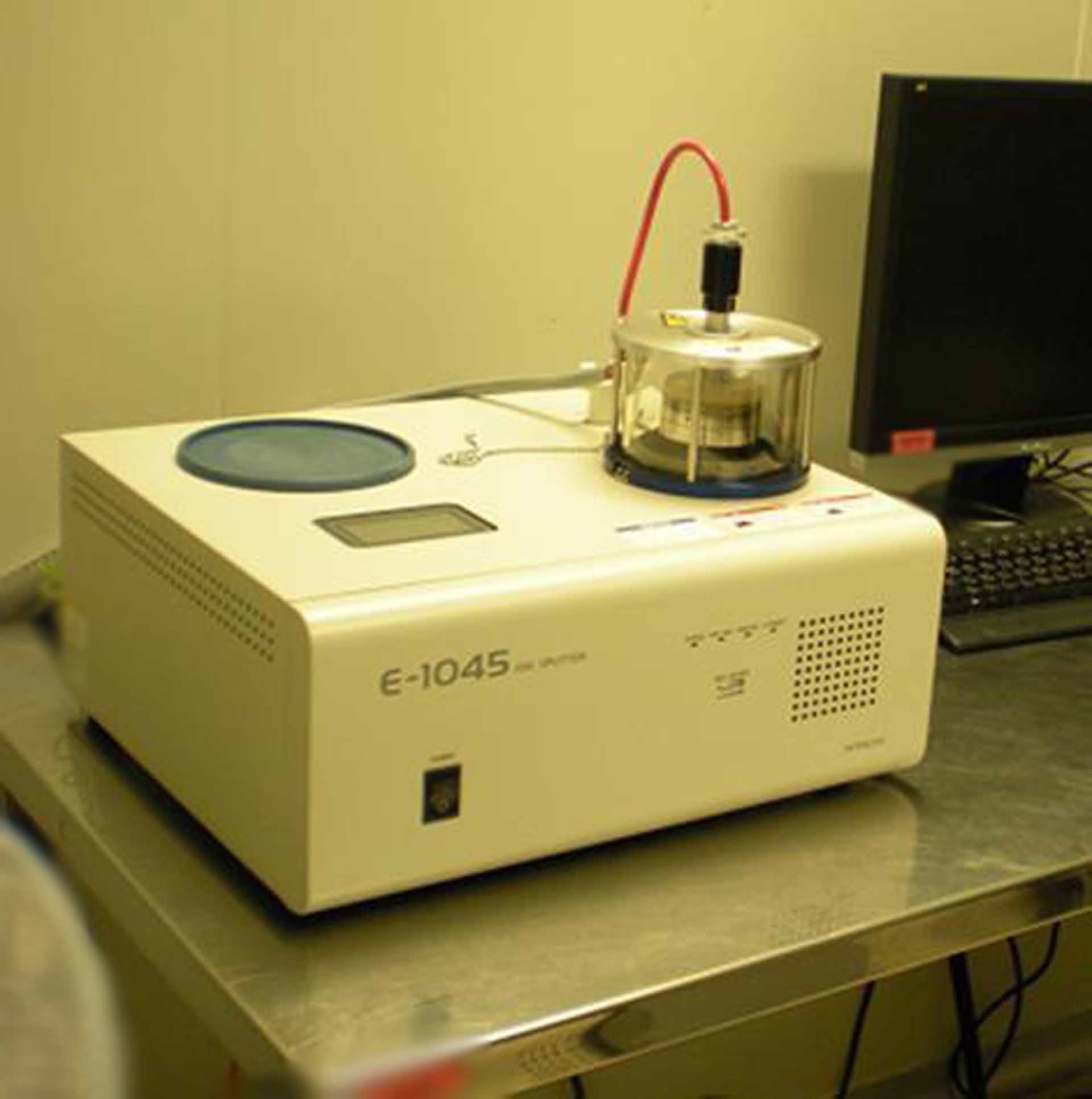
Ion coater rotary pump
LN2
UPS
EDS
Control HUB
EDS Controller
Operating system: Windows XP
Baking tool
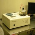
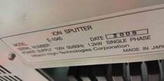
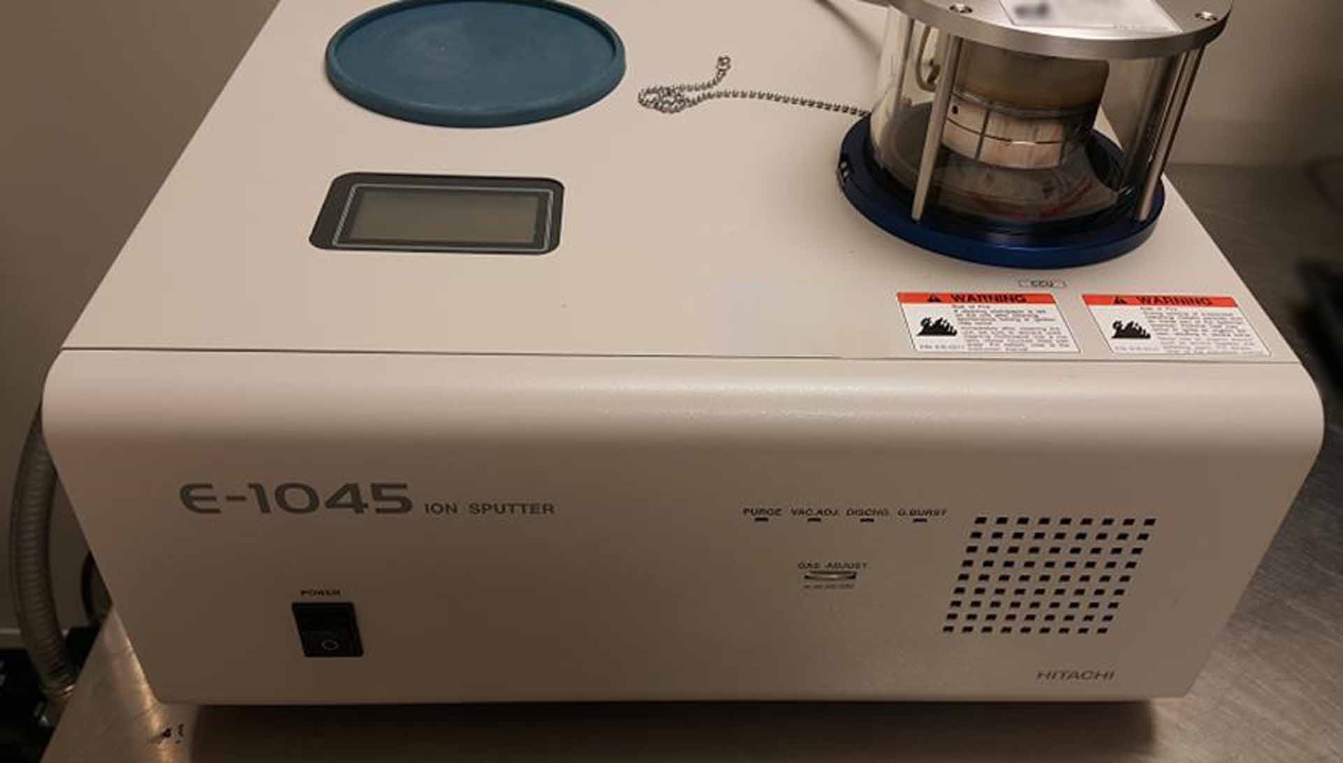
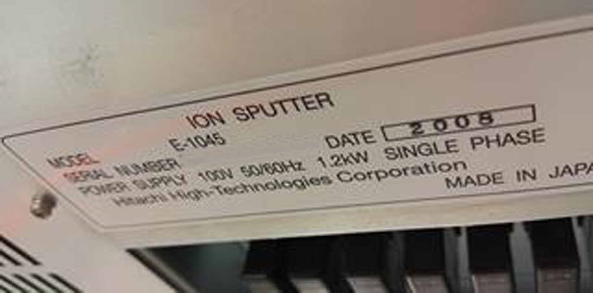
Ion coater
HP Office Jet Pro C8194A Printer
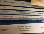
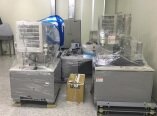
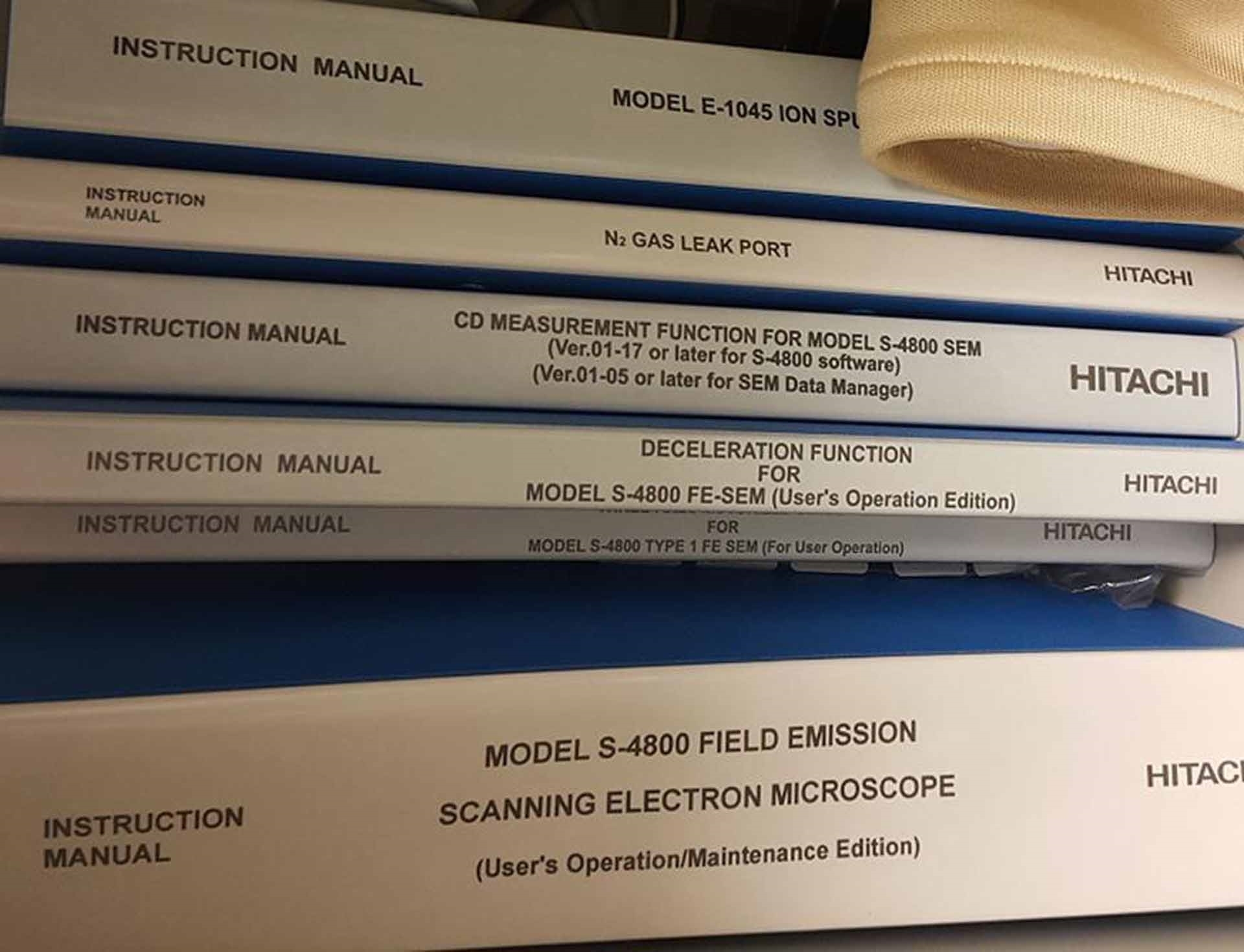
Manuals included
4 kVA For voltage other than 100V AC
Power requirements: 100V AC (±10% ), Single phase, 50/60 Hz
2008 vintage.
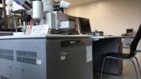
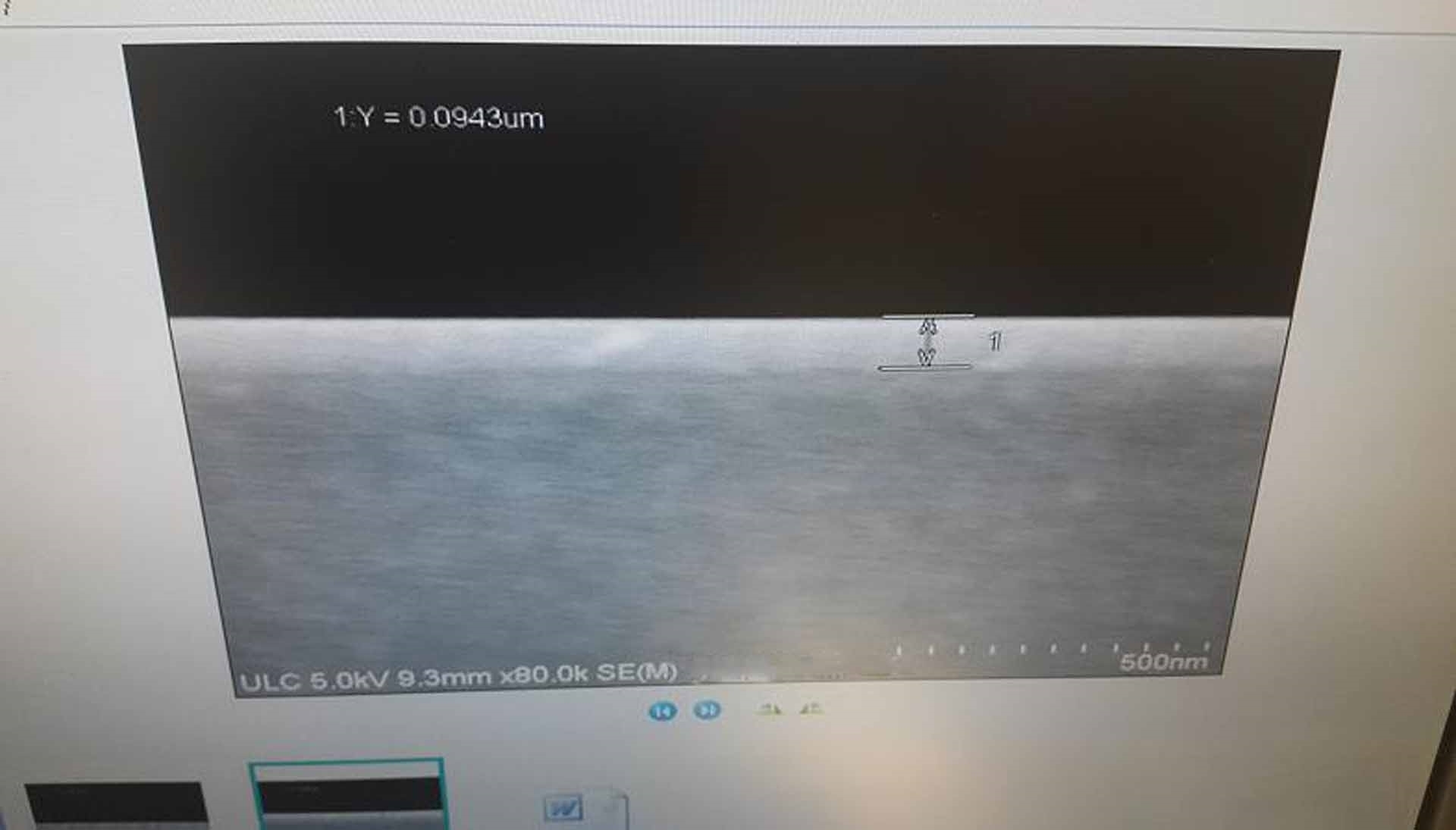
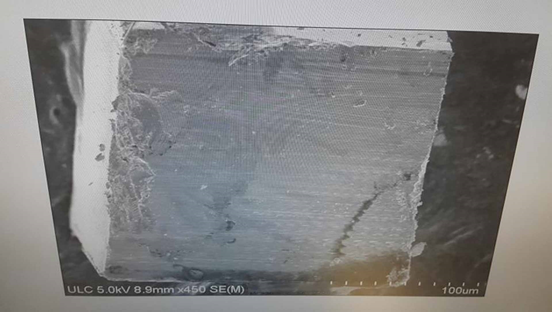
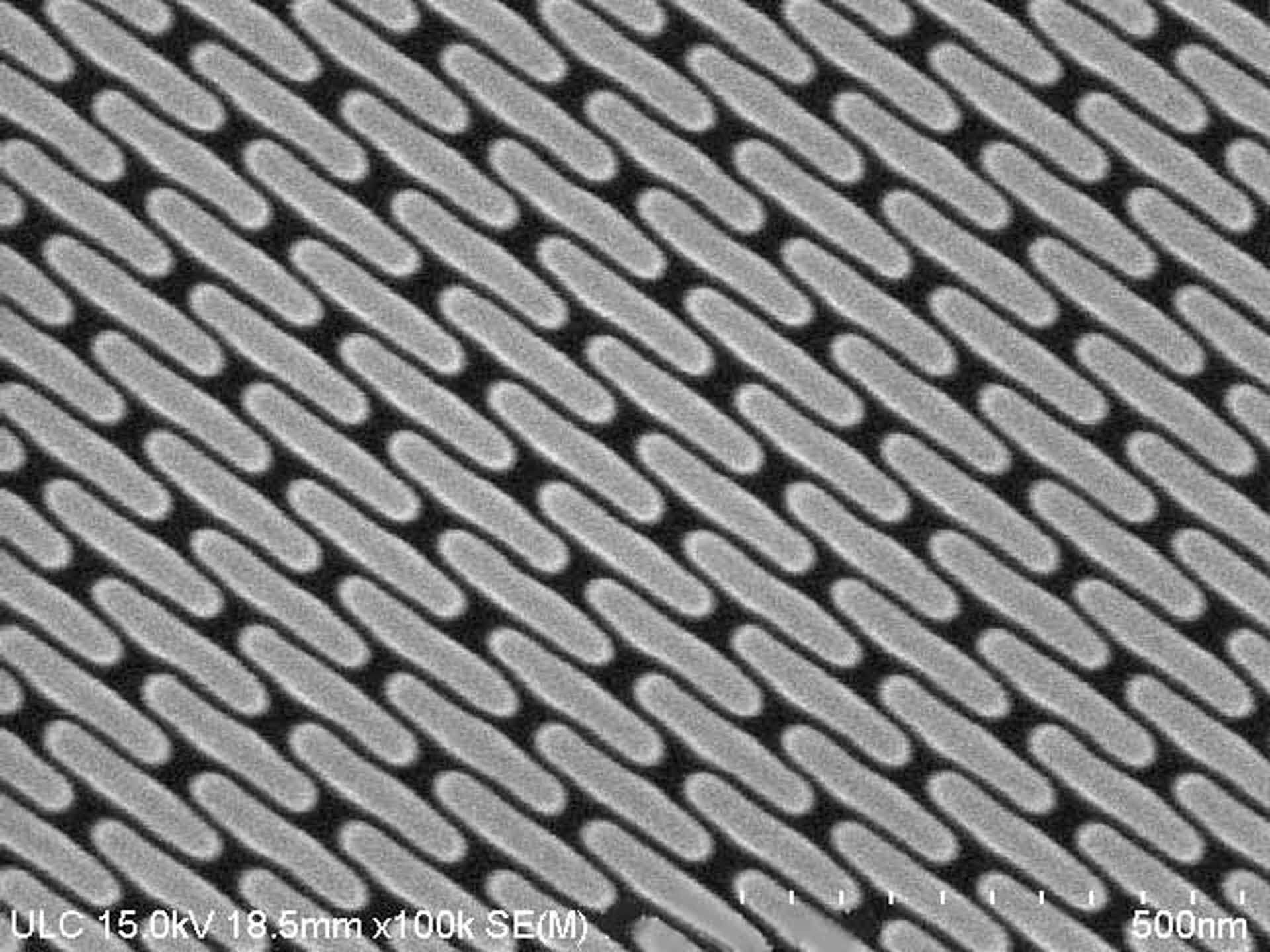
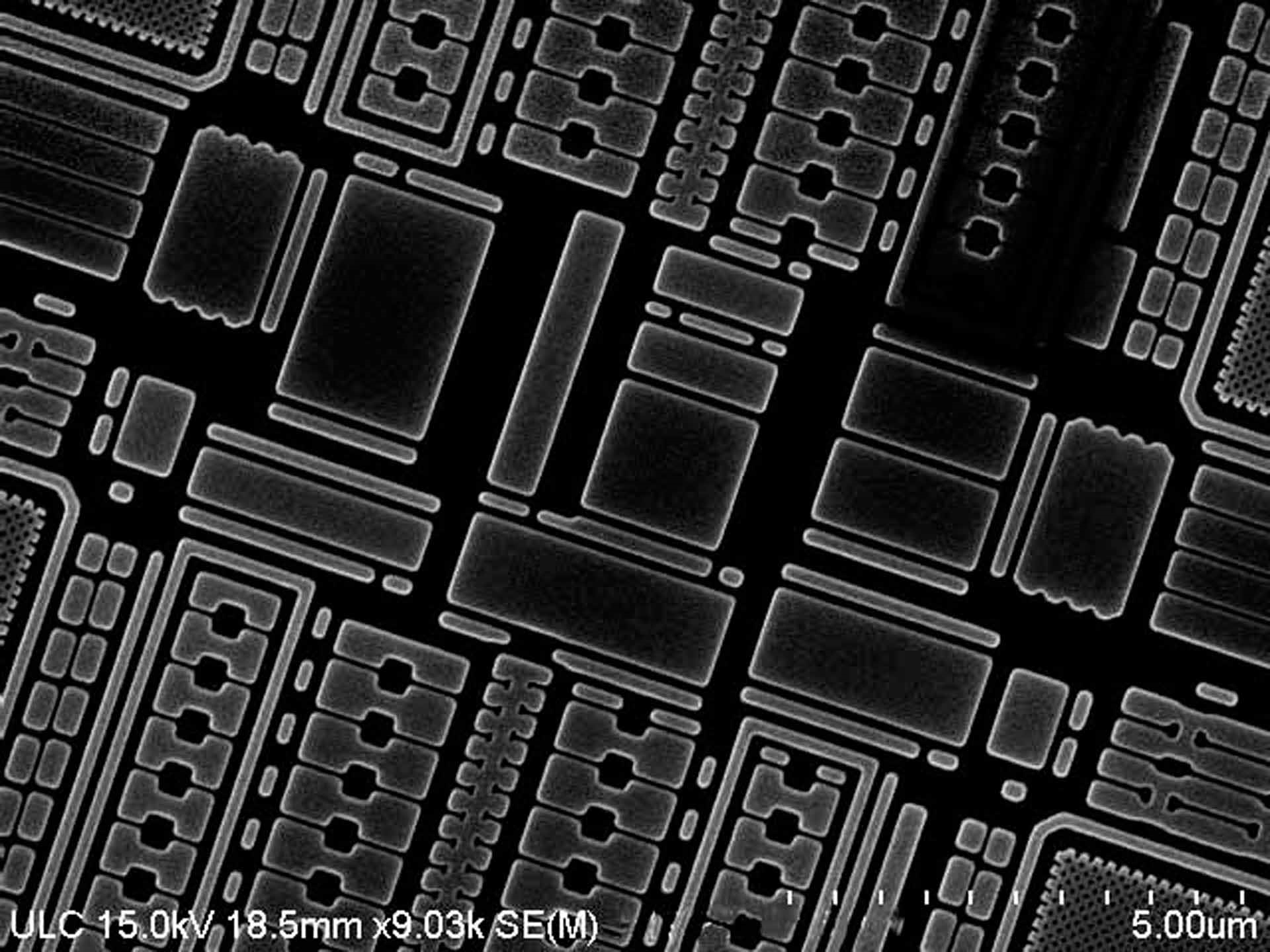
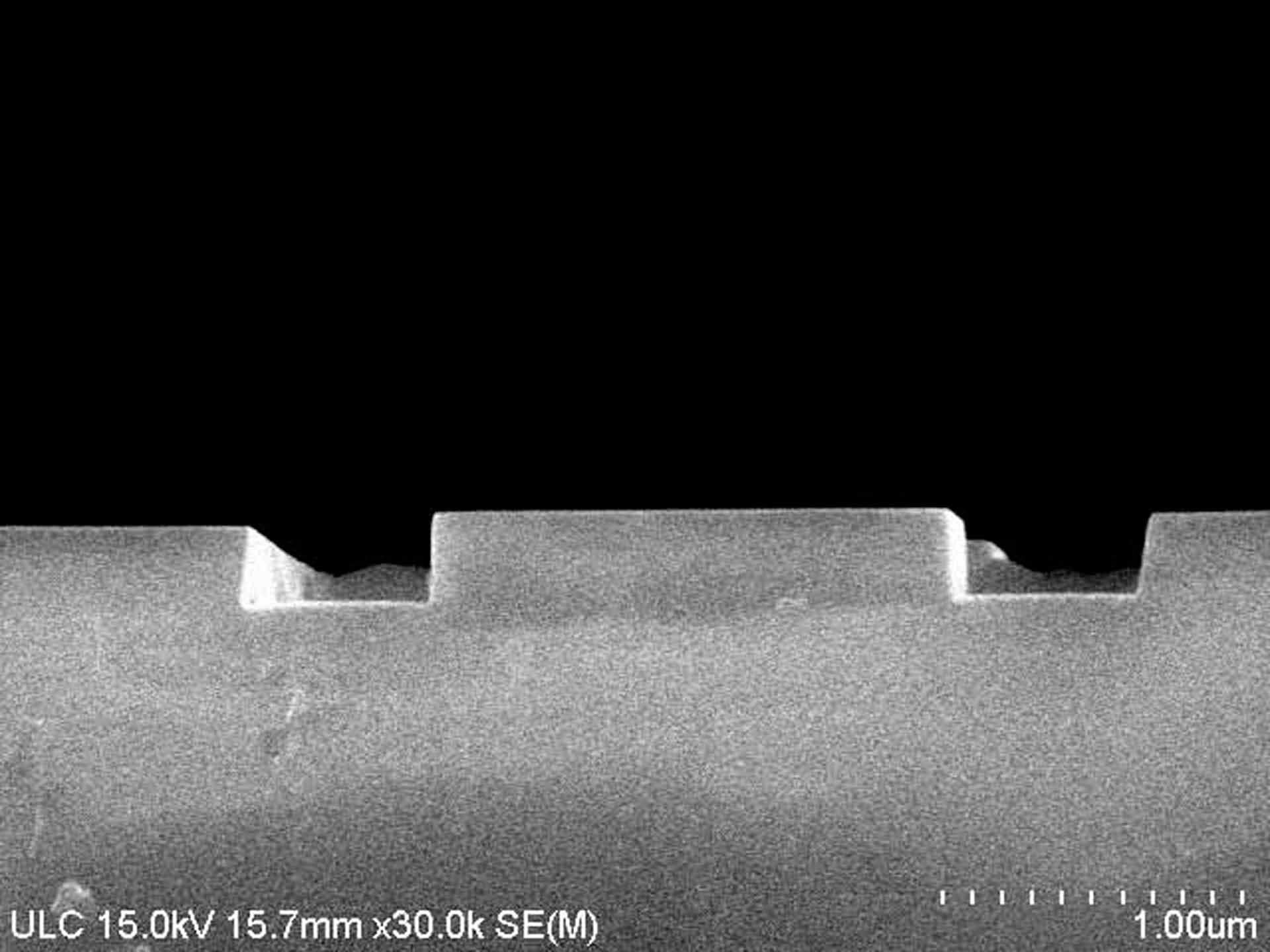
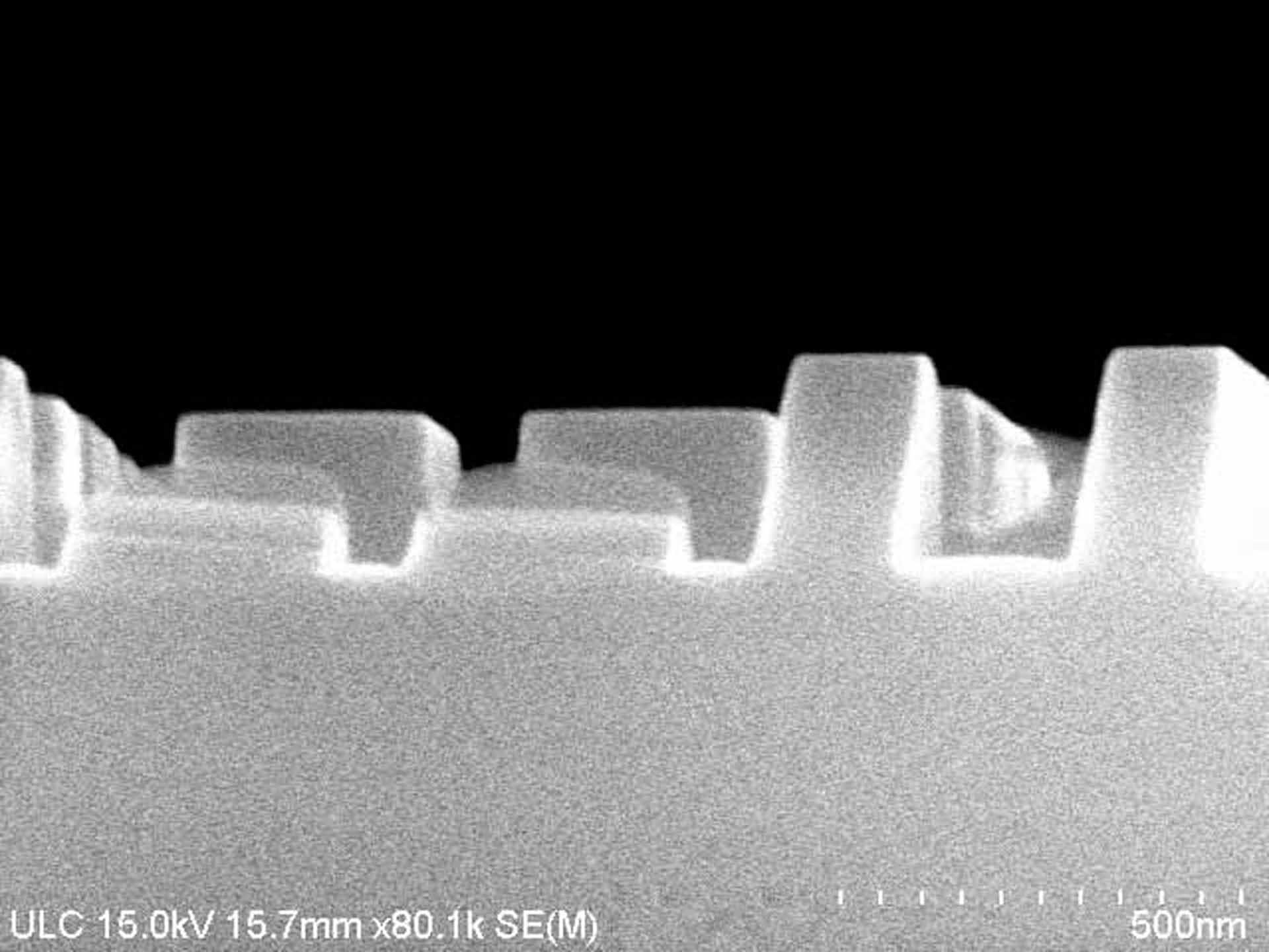
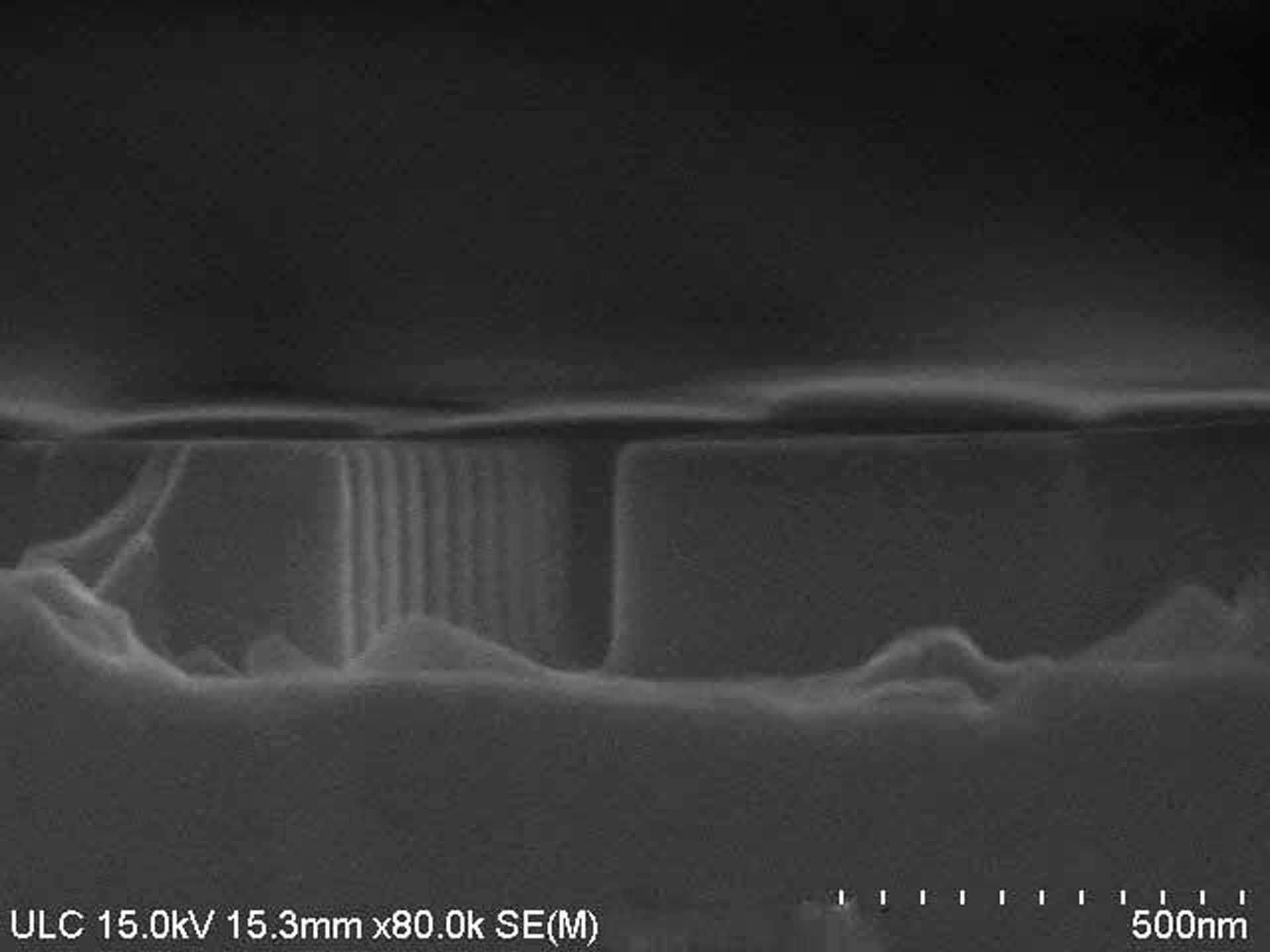
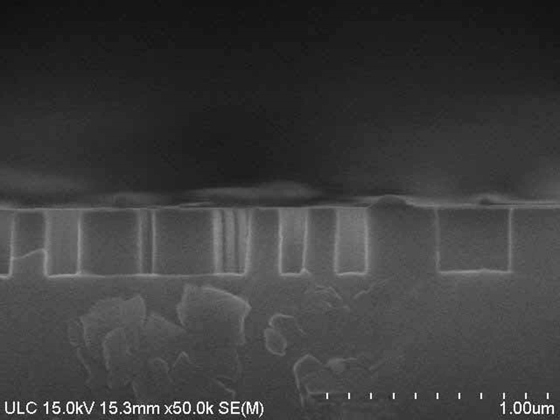
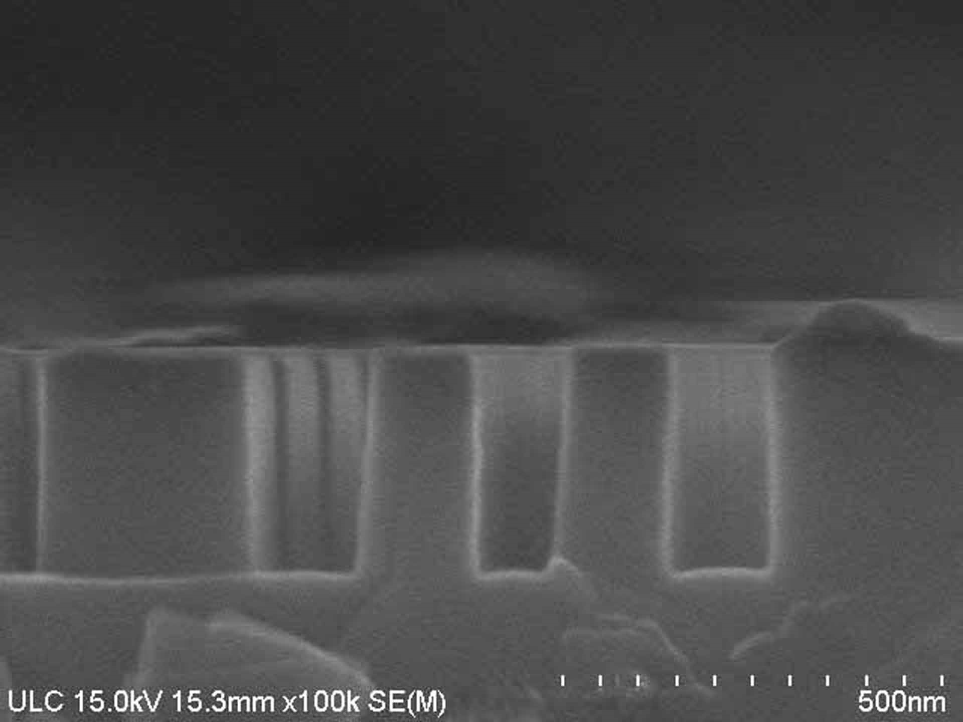
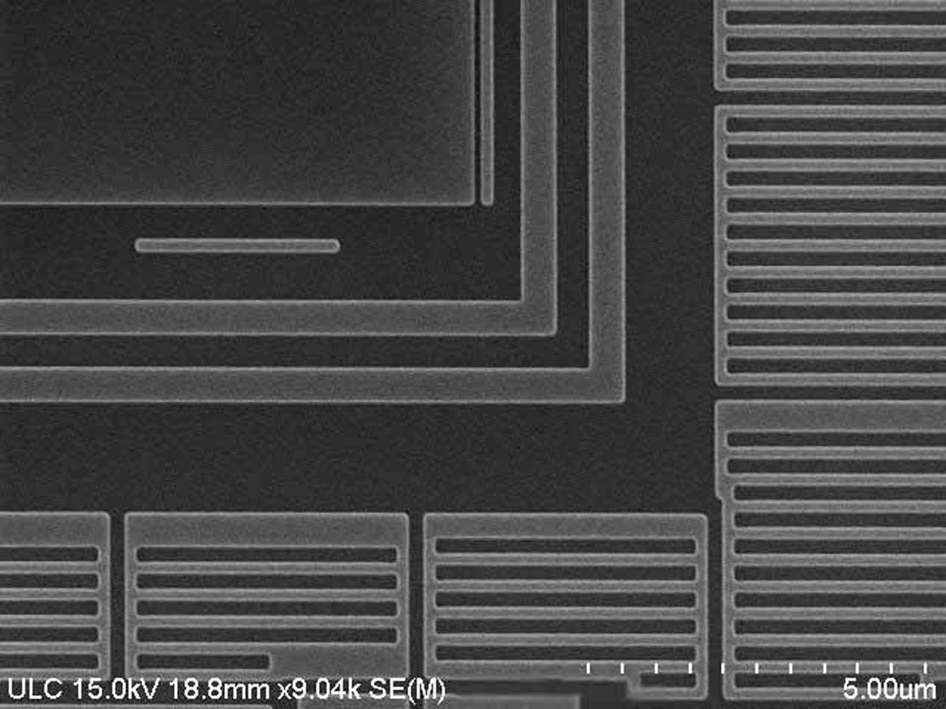
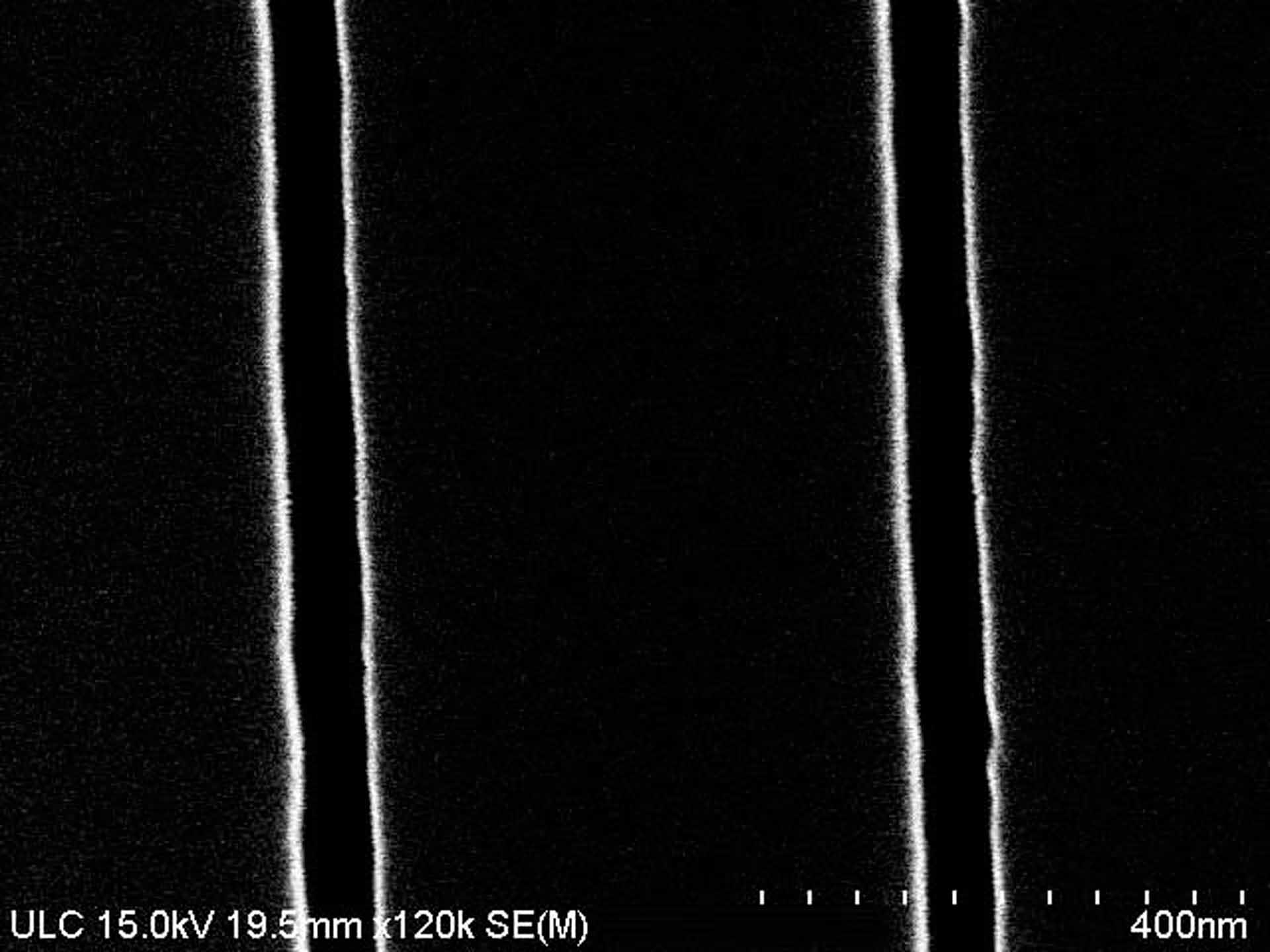
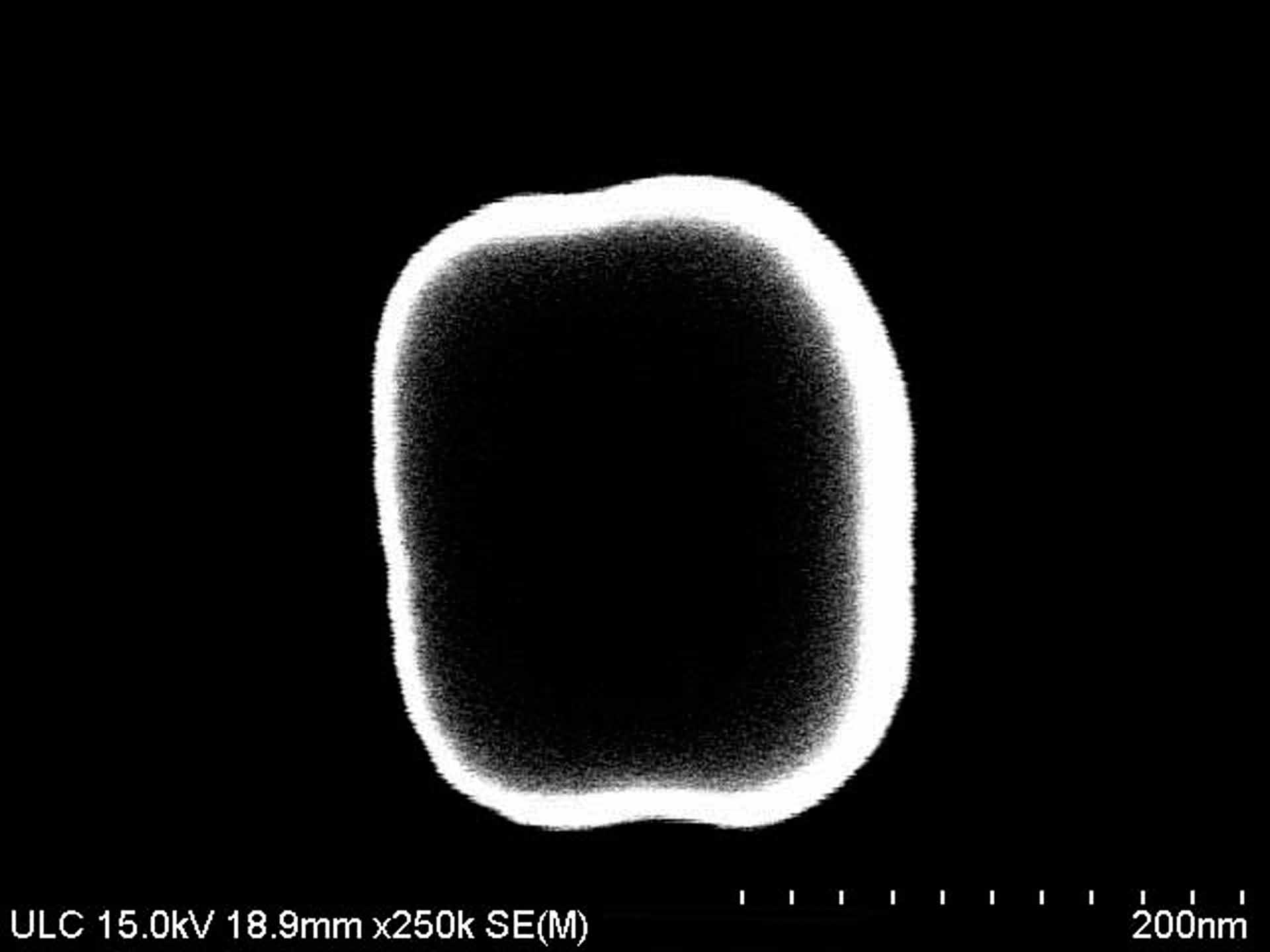
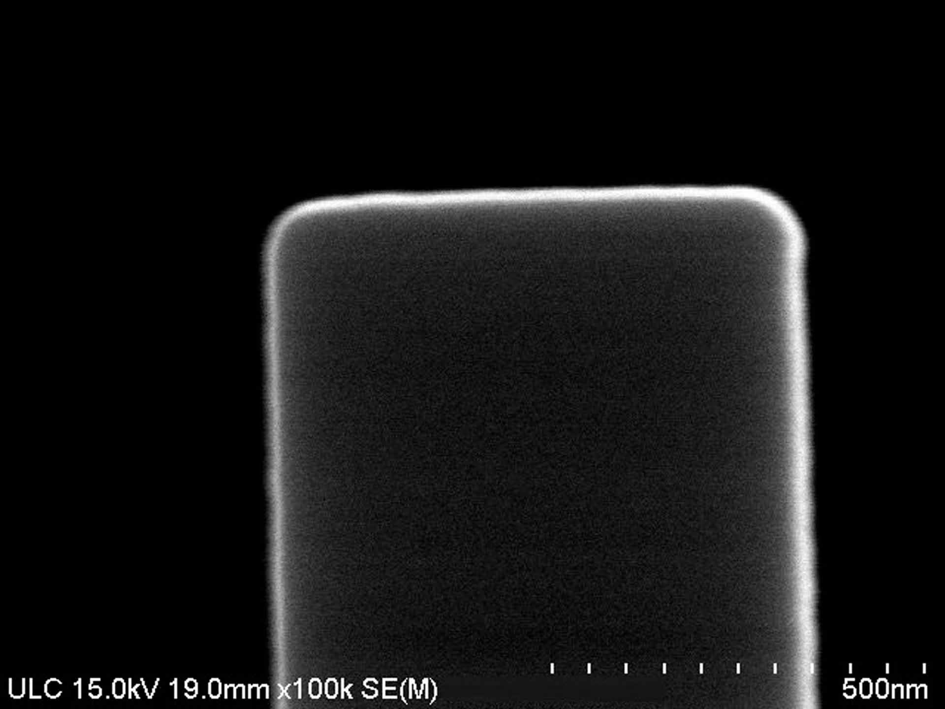
HITACHI S-4800은 뛰어난 이미지 품질과 다양한 분석 기능을 모두 제공하는 SEM (Scanning Electron Microscope) 입니다. 이 고급 도구는 재료 및 생명 과학 조사, 표본 분석, 실패 분석, 단면화 (cross-sectioning) 및 표면 측정과 같은 다양한 연구 및 산업 응용 분야를 위해 설계되었습니다. HITACHI S 4800에는 텅스텐 필라멘트 전자 방출기, 가변 압력 (VP) 및 가변 압력 가변 온도 (VPVT) 기능이 장착되어 있습니다. VP (VP) 및 VPVT (VPVT) 모드를 사용하면 다른 압력 수준과 온도에서 표본을 볼 수 있으므로 더 깊고 정확한 특성화가 가능합니다. 또한, 현미경은 직경 140mm의 큰 감지 영역을 특징으로하며 SEI (Secondary Electron Image) 모드에서 최대 1 nm 포인트 해상도의 높은 해상도를 갖습니다. 또한, ALV (Advanced Low Vacuum Mode) 는 비전도 재료에서 SEM Image를 감지 할 수있는 기능을 제공합니다. S-4800의 유연한 설계는 BSE (backscattered electron) 이미징, EDX 원소 분석, 질적 및 정량적 원소 분석을위한 WDS 등 다양한 기술에 사용될 수 있습니다. 또한 스테레오 및 3D 이미징 기능은 표본의 포괄적 인 특성을 제공합니다. 고급 이미징 (Advanced Imaging) 에는 라이브 (Live) 및 흑백 (Monochrome) 디스플레이와 디지털 이미지 측정 도구가 포함되어 있어 내부 구조 및 서피스 특성을 정확하게 측정 및 지정할 수 있습니다. 또한, 대형 샘플 챔버 및 고 리프트 스테이지는 효율적인 샘플 처리 및 조작을 가능하게합니다. S 4800 은 고급 탐지기 (advanced detector) 와 전자열 업그레이드 (electron column upgrade), 자동 이미징 시스템 (automatic imaging system) 등 다양한 액세서리를 갖추고 있습니다. 또한, 고급 에너지 필터링은 에너지 분산 X- 선 신호 연구에 도움이됩니다. 전반적으로 HITACHI S-4800 은 다양한 연구/산업 애플리케이션의 요구 사항을 충족하도록 설계된 강력한 하이엔드 SEM 입니다. 다목적 설계를 통해 뛰어난 이미지 품질 (Image Quality) 과 고급 분석 (Advanced Analysis) 기능을 제공하여 사용자가 분석되는 표본을 정확하고 정확하게 볼 수 있습니다.
아직 리뷰가 없습니다
